

Volume 16, Issue 4 (July & August 2025)
BCN 2025, 16(4): 819-832 |
Back to browse issues page



Download citation:
BibTeX | RIS | EndNote | Medlars | ProCite | Reference Manager | RefWorks
Send citation to:


BibTeX | RIS | EndNote | Medlars | ProCite | Reference Manager | RefWorks
Send citation to:
Golmohammadi M, Mostavafinia A, Nazarnejad M M, Nadia Sharifi Z. Effects of Nigella sativa Nano-hydroalcoholic Extract on Neuronal Damages in the Hippocampus of Male Rats After Cerebral Ischemia/Reperfusion. BCN 2025; 16 (4) :819-832
URL: http://bcn.iums.ac.ir/article-1-3162-en.html
URL: http://bcn.iums.ac.ir/article-1-3162-en.html
Mahsa Golmohammadi1 

 , Atarodalsadat Mostavafinia1 , Mohammad Mahdi Nazarnejad1 , Zahra Nadia Sharifi *2
, Atarodalsadat Mostavafinia1 , Mohammad Mahdi Nazarnejad1 , Zahra Nadia Sharifi *2
1- Department of Anatomical Sciences & Cognitive Neuroscience, TeMS.C., Islamic Azad University, Tehran, Iran.
2- Department of Anatomical Sciences & Cognitive Neuroscience, TeMS.C., Islamic Azad University, Tehran, Iran. & Herbal Pharmacology Research Center, TeMS.C., Islamic Azad University, Tehran, Iran.
2- Department of Anatomical Sciences & Cognitive Neuroscience, TeMS.C., Islamic Azad University, Tehran, Iran. & Herbal Pharmacology Research Center, TeMS.C., Islamic Azad University, Tehran, Iran.
Keywords: Hydroalcoholic extract, Hippocampus, Ischemia-reperfusion, Nigella sativa, Nano-hydroalcoholic extract
Full-Text [PDF 2195 kb]
| Abstract (HTML)
DLS analysis
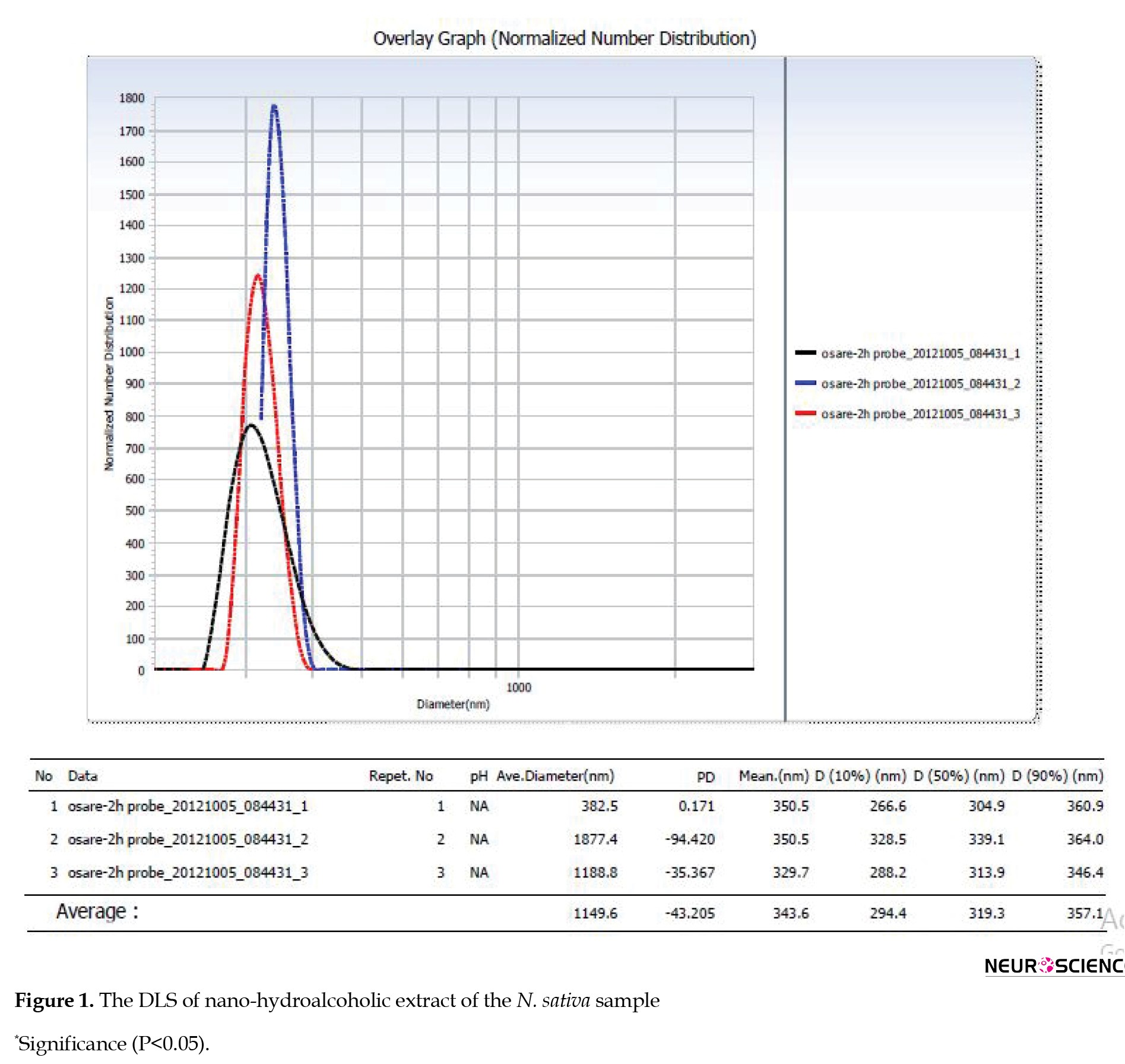
After the Nigella hydroalcoholic sample was prepared, DLS was employed to analyze particle size and distribution, and particle dispersion was examined using diagrams. The dispersion was between 294.4 and 357.1 nm, and a single peak diagram was obtained. The diameter of this diagram increases intensively when the size of particles exceeds 90 nm. If there are smaller particles with lower concentrations in the suspensions, they sometimes fade from the sensors, resulting in no intensity being reported. Following the stability test in 4 different environments, with one test conducted after one month and another after three months, the results remained consistent with the initial findings.
Investigation of particle size by zeta potential
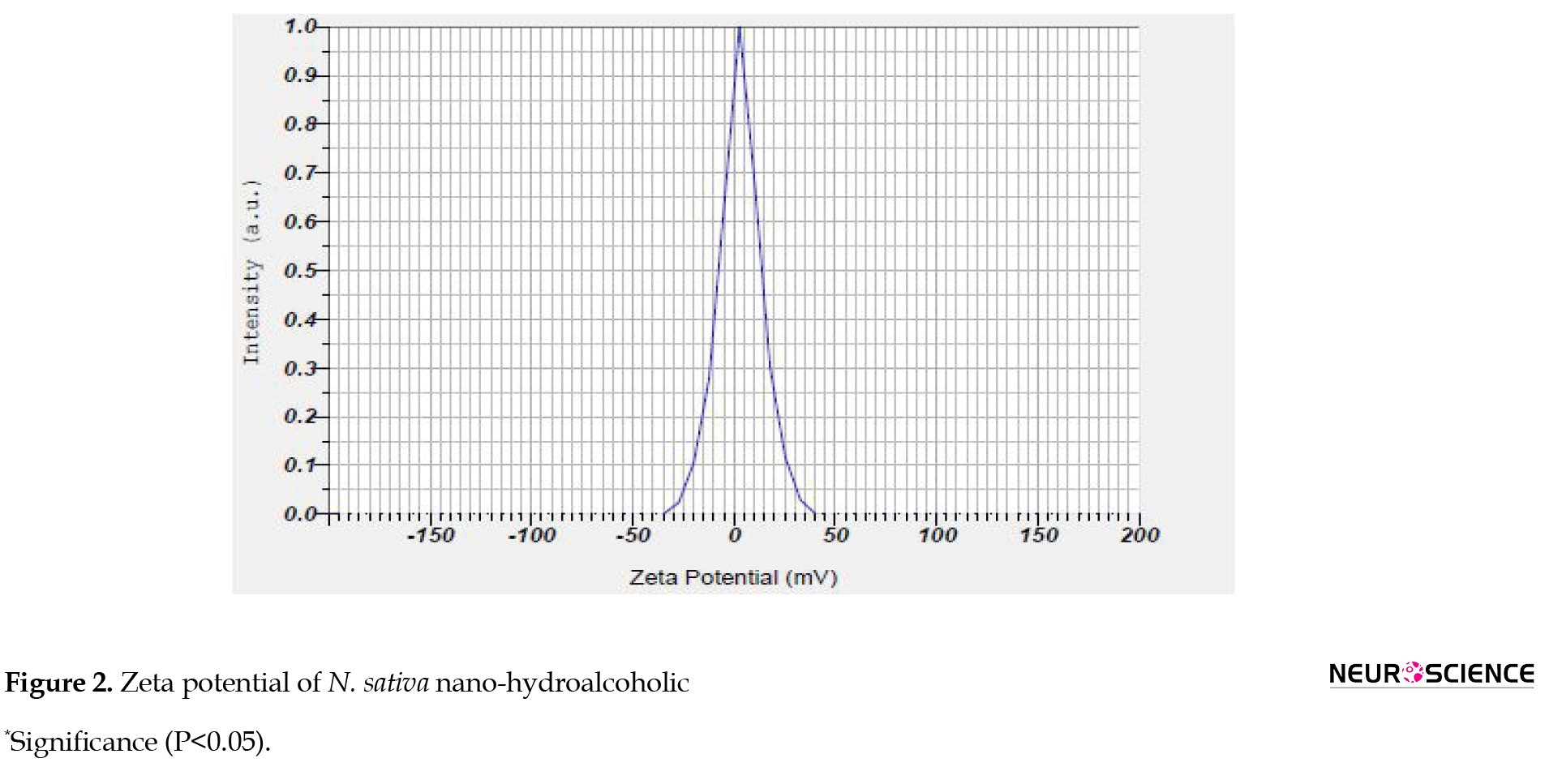
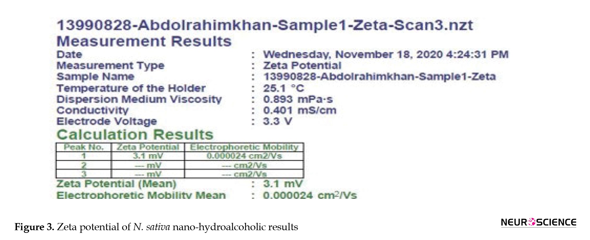
The results of the zeta potential of N. sativa nano-hydroalcoholic extract are as follows (Figures 2 and 3).
Zeta potential analysis
Most fluids contain cations and anions, which are atoms with positive and negative charges. When the loaded particles are suspended in a liquid, the opposing load ions are absorbed into the particulate matter. It means that samples with negative loads attract positive ions from the fluid, and conversely, samples with a positive charge attract negative ions from the fluid. The ions near the surface of the particle are highly absorbed while the farther ions will have a loose bond, called the intrusion layer. Zeta potential ranges from -10 to +10, indicating the stability of the sample (Figure 4).
Behavioral test results
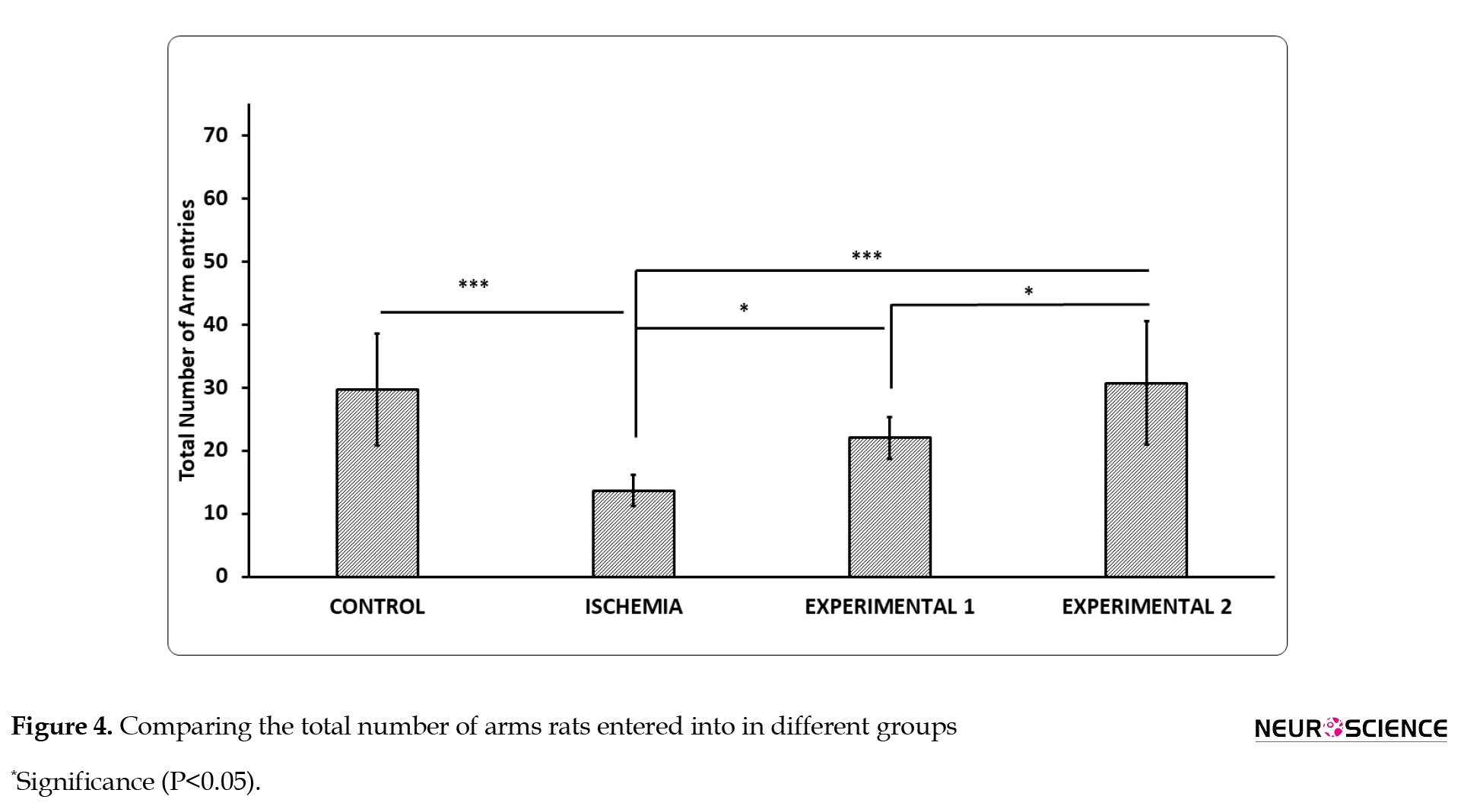
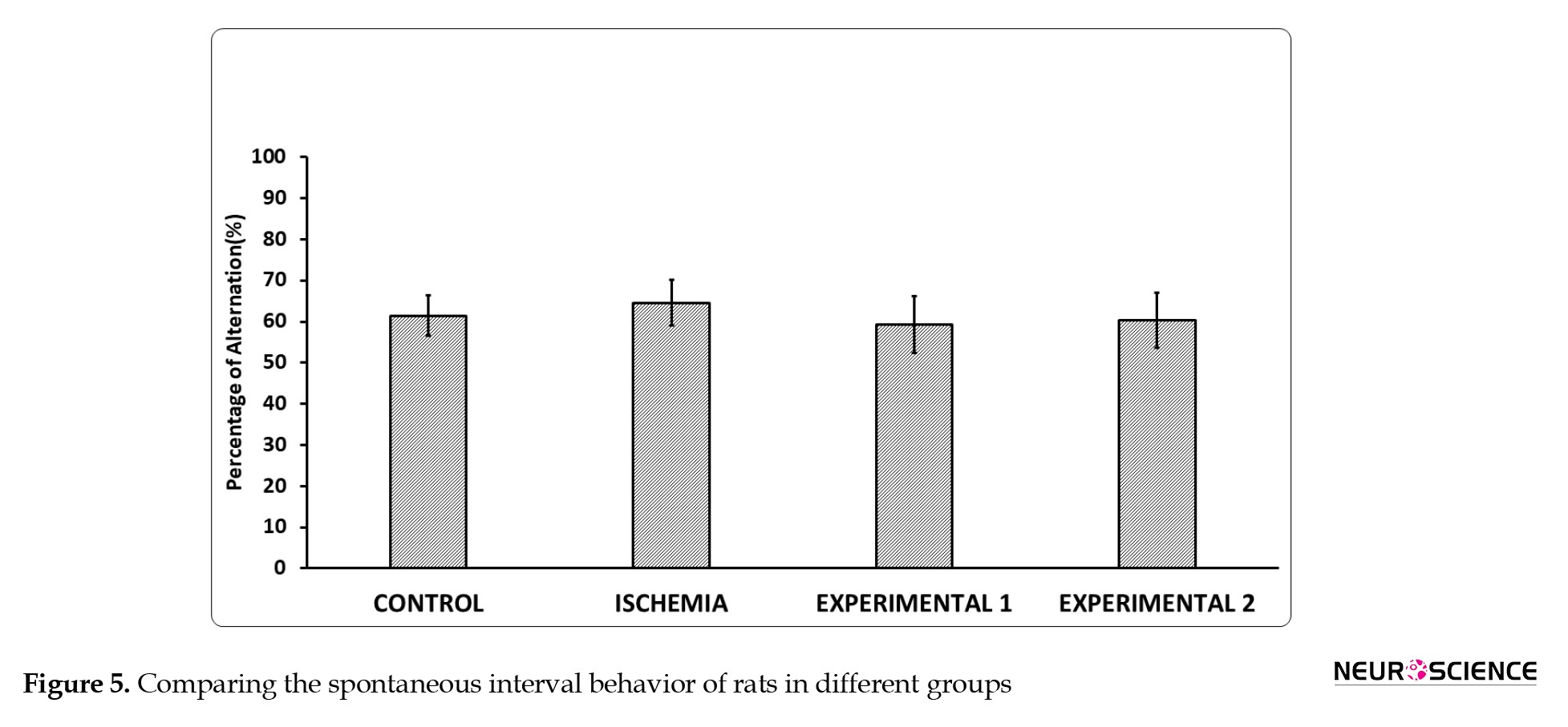
The results of the Y-maze test revealed that ischemia remarkably decreased the kinetic ability of test subjects. The aggregate of suitable feedback in the I/R group considerably declined in comparison with the control group. Furthermore, the longer the time of ischemia occurred, the worse the viciousness of neurological function was. Compared with the I/R group, the nano-hydroalcoholic extract of black seed recipient group showed significantly increased memory functions after I/R injury (Figures 4 and 5).
Results of the weight of rats in different groups
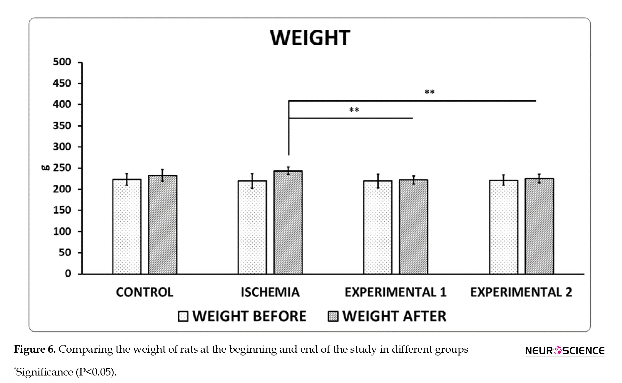
As a result of measuring the weight of rats before and after induction of ischemia and drug injection, it was observed that after surgery and during 14 days, when the experimental groups 1 and 2 were receiving the drug, one-way ANOVA test and LSD test showed a significant difference between ischemia group and experimental groups 1 and 2 (Figure 6).
Results of real-time PCR
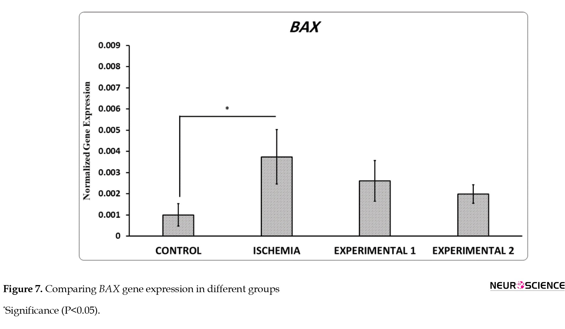
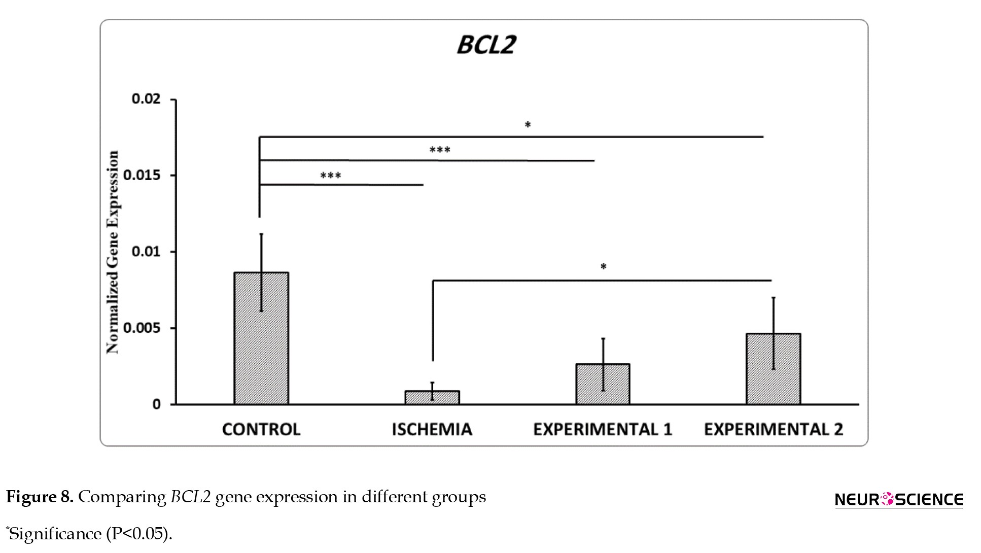
The relative expression of BAX in the hippocampus of the I/R group was considerably higher than that of the control group, whereas the relative expression of BCL-2 mRNA was substantially lower. (Figures 7 and 8).
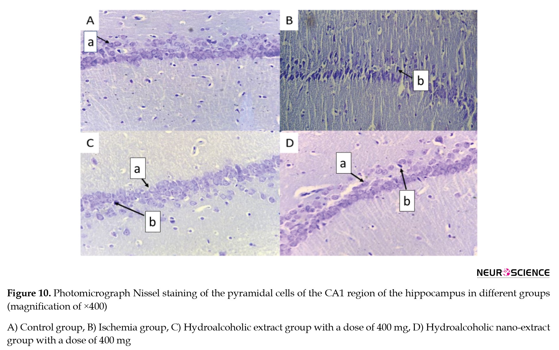
Most of the pyramidal cells were healthy. Note that a denotes healthy pyramidal cells, and b refers to degenerated pyramidal cells.
Results of stereological evaluation of the volume of the CA1 region of the hippocampus
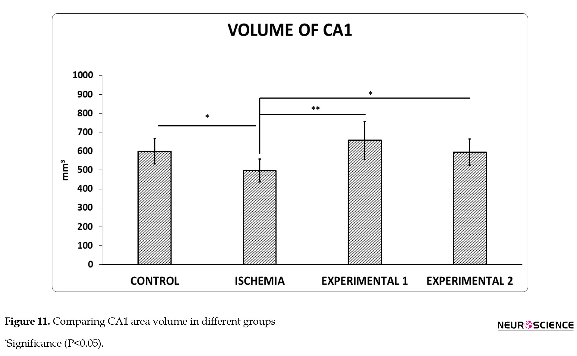
The results of calculating the volume of the hippocampus CA1 region of 6 histological slides with H&E staining for each rat and a total of 6 rats in each group were observed. The volume in the ischemia group decreased significantly compared to the control group. Furthermore, the volume of the hippocampus CA1 area by injection of hydroalcoholic extract increased significantly compared to the injection of N. sativa nano-hydroalcoholic extract (Figure 11).
4. Discussion
Our findings showed that cerebral ischemia/reperfusion for 20 minutes caused delayed death of pyramidal cells in the CA1 region.
Ischemia-reperfusion injury is a pathological condition that has both local and systemic effects. The key element during ischemia is cell energy depletion, and the interplay of oxidative and microcirculatory stress, as well as inflammation and apoptosis, takes place during reperfusion (Soares et al., 2019).
Following ischemia, hippocampus may be affected by programmed death of cells. Oxidative stress caused by reactive oxygen species (ROS) production contributes to the programmed death of cells (Itō, 1984). Increased ROS levels inside the cell can lead to DNA and intracellular protein damage through oxidation, resulting in cell death.
In response to global cerebral ischemia, CA1 neurons in the hippocampus are particularly susceptible and experience selective, delayed degeneration. These pyramidal neurons play a crucial role in spatial learning and memory, and their degeneration causes learning and memory problems (Xu et al., 2021; Zuo et al., 2015).
Antiinflammatory and antioxidant medications should be investigated to provide additional treatment options that can help prevent or reduce these injuries, as current drugs have limited efficacy and offer little to no benefit to patients (Mucha et al., 2021). Antioxidants are compounds that can significantly slow down the oxidative reaction (Molitoris & Wagner, 1996). So far, many natural and synthetic antioxidants have been introduced for the treatment or prevention of patients associated with free radicals (Farahmand & Tajali, 2016). Medicinal plants always have an instrumental role in the management of global health care and are considered the best treatment for diseases (Dehpour et al., 2011). N. sativa is a medicinal plant that is highly consumed. In recent years, further attention has been paid to the medicinal and biological properties of N. sativa.
One of N. sativa’s possible qualities is the ability of one or more of its components to minimize tissue damage following ischemic/reperfusion injury due to antioxidant activity (Bayrak et al., 2008; Erşahin et al., 2011).
It has been reported that administration of antioxidant agents before local brain ischemia in laboratory studies protects against ischemic brain lesion (Lipton., 1999). Based on a review of the literature, thymoquinone, which is the most important extract of N. sativa, has many pharmacological properties, mostly related to its antioxidant properties. In this regard, Hosseinzadeh et al. (2007) reported that the administration of Nigella oils in the global ischemic model of the brain has reduced the amount of lipid peroxidation. Their findings can support the role of antioxidant properties in the protective effects of N. sativa in cerebral ischemia. Ziyyat et al. (1997) investigated the neuroprotective effects of thymoquinone on transient cerebral ischemia on the hippocampus in rats. Thymoquinone reduces malondialdehyde levels, increases reduced glutathione, catalase, and superoxide dismutase activity until it reaches its normal level. Thymoquinone is a promising factor in healing the damages caused by the destruction of neural cells (neurodegeneration), such as cerebral ischemia. N. sativa also enhances the activity of antioxidant enzymes such as superoxide dismutase and glutathione peroxidase, and with its antioxidant properties, reduces lipid peroxidation in biological membranes (Butt & Sultan, 2010). It is known that thymoquinone and its active metabolite thymohydroquinone can prevent lipid peroxidation by sweeping superoxide, radical hydroxyl, and single molecular oxygen (Kanter, 2008). In addition, thymoquinone can inhibit arachidonic acid metabolism and restrict ischemic brain damage caused by inflammation of cyclooxygenase and lipoxygenase pathways (El Mezayen et al., 2006).
Nanoparticle-based drug delivery systems have demonstrated higher potential in treating ischemic stroke and potentially other neurological disorders. Joachim et al. showed that the gelatin of osteopontin nanoparticles in a rat ischemic stroke model augments nerve protection in the nose (Joachim et al., 2014). Nagai et al. showed that intravenous administration of silvestazole nanoparticles improves acute ischemic damage in the brain following the ischemic/reperfusion (Nagai et al. 2015). According to the previous studies and the results of the current research concerning the function of N. sativa plant and its role in preventing the death of neural cells, it can be said that the hydroalcoholic extract and nano-hydroalcoholic extract of N. sativa can exert its protective effect on ischemia/reperfusion brain damages, which can be attributed to the properties of oxygen free radicalization of this plant. Our results showed a significant decrease in BAX expression and an increase in BCL-2 expression following treatment with [compound/extract], suggesting a shift toward anti-apoptotic signaling. This BAX/BCL-2 modulation indicates that the therapy may protect against ischemia-induced neuronal apoptosis by promoting cell survival pathways. Also, the N. sativa plant in nano form has more efficacy and higher adsorption than the hydroalcoholic extract. Still, since the role of other compounds in these plants, which has not been studied, and the role of other mechanisms, such as inhibition of inflammatory pathways, and also the possibility of a cumulative effect of these compounds, is not known, this issue needs further research and studies.
5. Conclusion
This research revealed that hydroalcoholic extract and nano-hydroalcoholic extract containing N. sativa, by preventing morphological changes and resulting cell death, have an effective role on neurons in the CA1 region of the hippocampus. Therefore, it seems that hydroalcoholic extract and nano-hydroalcoholic extract containing N. sativa through antiinflammatory and antioxidant mechanisms can be considered as a treatment candidate for ischemic brain damage.
Ethical Considerations
This study was approved by the Ethics Committee of Tehran Medical Sciences Branch, Islamic Azad University, Tehran, Iran (Code: IR.IAU.TMU.REC.1399.515). The ARRIVE ethical criteria were followed in all of the trials. During the research, the Guide for the Care and Use of Laboratory Animals (NIH Publication No. 86-23) was also followed.
Funding
This research did not receive any grant from funding agencies in the public, commercial, or non-profit sectors.
Authors' contributions
Conceptualization, supervision, data analysis, interpretation, review and editing: Zahra Nadia Sharifi and Atarodalsadat Mostavafinia; Methodology and experimental design: Mahsa Golmohammadi and Atarodalsadat Mostavafinia; Data collection and laboratory work: Mahsa Golmohammadi and Mohammad Mahdi Nazarnejad; Original draft preparation: Mohammad Mahdi Nazarnejad; Final approval: All authors.
Conflict of interest
The authors declared no conflict of interest.
Acknowledgments
The authors are grateful to the Herbal Pharmacology Research Center, Tehran Medical Sciences Branch, Islamic Azad University, Tehran, Iran, and all of the personnel For their direction and support,
References
Antonsson, B., & Martinou, J. C. (2000). The Bcl-2 protein family. Experimental Cell Research, 256(1), 50-57. [DOI:10.1006/excr.2000.4839] [PMID]
Bayrak, O., Bavbek, N., Karatas, O. F., Bayrak, R., Catal, F., & Cimentepe, E., et al. (2008). Nigella sativa protects against ischaemia/reperfusion injury in rat kidneys. Nephrology, Dialysis, Transplantation : Official Publication of the European Dialysis and Transplant Association - European Renal Association, 23(7), 2206–2212. [DOI:10.1093/ndt/gfm953] [PMID]
Butt, M. S., & Sultan, M. T. (2010). Nigella sativa: Reduces the risk of various maladies. Critical Reviews in Food Science and Nutrition, 50(7), 654-665. [DOI:10.1080/10408390902768797] [PMID]
Dehpour, A. A., Ebrahimzadeh, M. A., Roudgar, A., Nabavi, S.F., & Nabavi, S. M. (2011). Antioxidant and antibacterial activity of Consolida orientalis. World Acad Sci Eng Technol, 73:162-6. [Link]
Dellu, F., Mayo, W., Cherkaoui, J., Le Moal, M., & Simon, H. (1992). A two-trial memory task with automated recording: Study in young and aged rats. Brain Research, 588(1), 132-139. [DOI:10.1016/0006-8993(92)91352-F] [PMID]
Erşahin, M., Toklu, H. Z., Akakin, D., Yuksel, M., Yeğen, B. Ç., & Sener, G. (2011). The effects of Nigella sativa against oxidative injury in a rat model of subarachnoid hemorrhage. Acta Neurochirurgica, 153(2), 333-341. [DOI:10.1007/s00701-010-0853-9] [PMID]
El Mezayen, R., El Gazzar, M., Nicolls, M. R., Marecki, J. C., Dreskin, S. C., & Nomiyama, H. (2006). Effect of thymoquinone on cyclooxygenase expression and prostaglandin production in a mouse model of allergic airway inflammation. Immunology Letters, 106(1), 72-81. [DOI:10.1016/j.imlet.2006.04.012] [PMID]
Farahmand, M., & Tajali, M. (2016). [The effect of Salvia rhytidea extract on the Number of cells of different Layers of cerebellar cortex following ischemia reperfusion in rats (Persian)]. Armaghane Danesh, 21(6), 536-551. [Link]
Graham, S. H., & Chen, J. (2001). Programmed cell death in cerebral ischemia. Journal of Cerebral Blood Flow & Metabolism, 21(2), 99-109. [DOI:10.1097/00004647-200102000-00001] [PMID]
Hajhashemi, V., Ghannadi, A., & Jafarabadi, H. (2004). Black cumin seed essential oil, as a potent analgesic and antiinflammatory drug. Phytotherapy Research: An International Journal Devoted to Pharmacological and Toxicological Evaluation of Natural Product Derivatives, 18(3), 195-199. [DOI:10.1002/ptr.1390] [PMID]
Hosseinzadeh, H., Parvardeh, S., Asl, M. N., Sadeghnia, H. R., & Ziaee, T. (2007). Effect of thymoquinone and Nigella sativa seeds oil on lipid peroxidation level during global cerebral ischemia-reperfusion injury in rat hippocampus. Phytomedicine, 14(9), 621-627. [DOI:10.1016/j.phymed.2006.12.005] [PMID]
Itō, M. (1984). The cerebellum and neural control. New York: Raven Press. [Link]
Jiao, L., Zhang, J., Li, Z., Liu, H., Chen, Y., & Xu, S. (2011). Edaravone alleviates delayed neuronal death and long-dated cognitive dysfunction of hippocampus after transient focal ischemia in Wistar rat brains. Neuroscience, 182, 177-183. [DOI:10.1016/j.neuroscience.2011.01.017] [PMID]
Joachim, E., Kim, I. D., Jin, Y., Kim, K. K., Lee, J. K., & Choi, H. (2014). Gelatin nanoparticles enhance the neuroprotective effects of intranasally administered osteopontin in rat ischemic stroke model. Drug Delivery and Translational Research, 4(5-6), 395-399. [DOI:10.1007/s13346-014-0208-9] [PMID]
Kaplan, D. R., & Miller, F. D. (2000). Neurotrophin signal transduction in the nervous system. Current Opinion in Neurobiology, 10(3), 381-391. [DOI:10.1016/S0959-4388(00)00092-1] [PMID]
Kanter, M., Coskun, O., & Uysal, H. (2006). The antioxidative and antihistaminic effect of Nigella sativa and its major constituent, thymoquinone on ethanol-induced gastric mucosal damage. Archives of Toxicology, 80(4), 217-224. [DOI:10.1007/s00204-005-0037-1] [PMID]
Kanter, M., Coskun, O., Kalayci, M., Buyukbas, S., & Cagavi, F. (2006). Neuroprotective effects of Nigella sativa on experimental spinal cord injury in rats. Human & Experimental Toxicology, 25(3), 127-133. [DOI:10.1191/0960327106ht608oa] [PMID]
Kanter, M. (2008). Nigella sativa and derived thymoquinone prevents hippocampal neurodegeneration after chronic toluene exposure in rats. Neurochemical Research, 33(3), 579-588. [DOI:10.1007/s11064-007-9481-z] [PMID]
Kirino, T., & Sano, K. (1984). Selective vulnarablility in the gerbil hippocampus following transient ischemia. Actaneuropathologica, 62(3), 201-208. [DOI: 10.1007/BF00691853] [PMID]
Longa, E. Z., Weinstein, P. R., Carlson, S., & Cummins, R. (1989). Reversible middle cerebral artery occlusion without craniectomy in rats. Stroke, 20(1), 84-91. [DOI:10.1161/01.STR.20.1.84] [PMID]
Lipton, P. (1999). Ischemic cell death in brain neurons. Physiological Reviews, 79(4), 1431–1568. [DOI:10.1152/physrev.1999.79.4.1431] [PMID]
Meddah, B., Ducroc, R., Faouzi, M. E. A., Eto, B., Mahraoui, L., & Benhaddou-Andaloussi, A., et al. (2009). Nigella sativa inhibits intestinal glucose absorption and improves glucose tolerance in rats. Journal of Ethnopharmacology, 121(3), 419-424. [DOI:10.1016/j.jep.2008.10.040] [PMID]
Mucha, P., Skoczyńska, A., Małecka, M., Hikisz, P., & Budzisz, E. (2021). Overview of the Antioxidant and Anti-Inflammatory Activities of Selected Plant Compounds and Their Metal Ions Complexes. Molecules (Basel, Switzerland), 26(16), 4886. [DOI:10.3390/molecules26164886] [PMID]
Molitoris, B. A., & Wagner, M. C. (1996). Surface membrane polarity of proximal tubular cells: alterations as a basis for malfunction. Kidney International, 49(6), 1592-1597. [DOI:10.1038/ki.1996.231] [PMID]
Nagai, N., Yoshioka, C., Ito, Y., Funakami, Y., Nishikawa, H., & Kawabata, A. (2015). Intravenous administration of cilostazol nanoparticles ameliorates acute ischemic stroke in a cerebral ischemia/reperfusion-induced injury model. International Journal of Molecular Sciences, 16(12), 29329-29344. [DOI:10.3390/ijms161226166] [PMID]
Peng, H., Li, Y. F., & Sun, S. G. (2003). Effects of Ginkgo biloba extract on acute cerebral ischemia in rats analyzed by magnetic resonance spectroscopy. Acta Pharmacologica Sinica, 24(5), 467-471. [PMID]
Soares, R. O., Losada, D. M., Jordani, M. C., Évora, P., & Castro-e-Silva, O. (2019). Ischemia/reperfusion injury revisited: an overview of the latest pharmacological strategies. International Journal of Molecular Sciences, 20(20), 5034. [DOI:10.3390/ijms20205034] [PMID]
Tanaka, H., Grooms, S. Y., Bennett, M. V., & Zukin, R. S. (2000). The AMPAR subunit GluR2: Still front and center-stage. Brain Research, 886(1-2), 190-207. [DOI:10.1016/S0006-8993(00)02951-6]
Tavakkoli, A., Ahmadi, A., Razavi, B. M., & Hosseinzadeh, H. (2017). Black seed (Nigella sativa) and its constituent thymoquinone as an antidote or a protective agent against natural or chemical toxicities. Iranian Journal of Pharmaceutical Research: IJPR, 16(Suppl), 2-23. [PMID]
Tekeoglu, I., Dogan, A., Ediz, L., Budancamanak, M., & Demirel, A. (2007). Effects of thymoquinone (volatile oil of black cumin) on rheumatoid arthritis in rat models. Phytotherapy Research: An International Journal Devoted to Pharmacological and Toxicological Evaluation of Natural Product Derivatives, 21(9), 895-897. [DOI:10.1002/ptr.2143] [PMID]
Xu, H., Wang, E., Chen, F., Xiao, J., & Wang, M. (2021). Neuroprotective phytochemicals in experimental ischemic stroke: mechanisms and potential clinical applications. Oxidative Medicine and Cellular Longevity, 2021, 6687386. [DOI:10.1155/2021/6687386] [PMID]
Yamashima, T., & Oikawa, S. (2009). The role of lysosomal rupture in neuronal death. Progress in Neurobiology, 89(4), 343-358. [DOI:10.1016/j.pneurobio.2009.09.003] [PMID]
Zuo, W., Zhang, W., Han, N., & Chen, N. H. (2015). Compound IMM-H004, a novel coumarin derivative, protects against CA1 cell loss and spatial learning impairments resulting from transient global ischemia. CNS Neuroscience & Therapeutics, 21(3), 280–288. [DOI:10.1111/cns.12364] [PMID]
Ziyyat, A., Legssyer, A., Mekhfi, H., Dassouli, A., Serhrouchni, M., & Benjelloun, W. (1997). Phytotherapy of hypertension and diabetes in oriental Morocco. Journal of Ethnopharmacology, 58(1), 45-54. [DOI:10.1016/S0378-8741(97)00077-9] [PMID]
Full-Text:
1. Introduction
Transient global ischemia in humans occurs due to cardiac arrest or cardiac surgery, which causes selective and delayed nerve death, especially pyramidal neurons in the hippocampus (Tanaka et al., 2000). The key structure of the memory system is the hippocampus, which is used to study phenomena related to ischemia. However, specific hippocampal subsets illustrate different sensitivities to ischemia/hypoxia. The most sensitive area of the hippocampus is CA1. Even if it is exposed to a short-term ischemic attack, the mechanisms of delayed neuronal death leading to a significant reduction of nerve cells occur in this structure (Jiao et al., 2011). It has been reported that CA2-3 segments are relatively resistant to ischemia. Long-term ischemia due to ischemic recurrence causes changes in CA2-3 neurons, but the reduction of neurons, which is evident in this part, is rarely shown (Kirino & Sano, 1984). Nerve death due to global ischemia occurs up to 2-4 days after ischemia in rats, and the fetus is not recognizable. The role of apoptotic death and necrosis in ischemia-induced nerve cell death remains controversial (Yamashima & Oikawa, 2009; Graham & Chen, 2001).
However, the underlying mechanisms of ischemic death are still unclear. The molecular events that determine the fate of neurons could be investigated due to the significant delay between attack and the onset of death. It has been shown that extracts of herbs preserve protection against ischemic damage to various organs such as the kidneys, heart, and brain (Peng et al., 2003). Apoptosis, or programmed cell death, plays a critical role in neurodegenerative conditions and ischemia-induced brain damage. Among the key regulators of apoptosis are the BCL-2 family proteins, which include both pro-apoptotic and anti-apoptotic members. BAX (Bcl-2-associated X protein) promotes apoptosis by facilitating mitochondrial outer membrane permeabilization, whereas BCL-2 (B-cell lymphoma 2) inhibits this process and promotes cell survival. The ratio of BAX to BCL-2 is often used as an indicator of a cell’s susceptibility to apoptosis. Changes in the expression of BAX and BCL-2 genes can indicate whether a treatment exerts pro-survival or pro-apoptotic effects on neurons.
Therefore, assessing these markers helps evaluate the neuroprotective potential of experimental treatments (Antonsson & Martinou, 2000). In the past few years, scientists have worked to identify the active compounds in herbal medications and to understand how they work (Meddah et al., 2009). Black seed, or Nigella sativa, a member of the Ranunculaceae family, is one of these herbal treatments that is used for a broad range of diseases, including bronchial asthma, headache, allergies, gastrointestinal problems, hypertension, obesity, and various types of cancer. N. sativa seeds can also help reduce depression and have immunostimulatory effects on various inflammatory and immunologic diseases, including arthritis, colitis, and experimental allergic encephalomyelitis, as well as in sensitized animals, asthma patients, and chemical warfare victims, due to its documented component (Kaplan & Miller, 2000). Recently, clinical and animal studies on therapeutic drugs of black seed extracts have revealed that these drugs include a variety of effects such as anti inflammation (Hajhashemi et al., 2004) and modulation of the immune system (Tekeoglu., 2007), antioxidant activities (Kanter et al., 2006), and protection from light (Kanter et al., 2006). Recently, considerable attention has been focused on plants containing natural products and agents that can mitigate the damage caused by free radicals. The current research aims to explore the effect of hydroalcoholic and nano-hydroalcoholic extracts containing N. sativa on neurons in the CA1 area of the hippocampus in an ischemia model of the Wistar rats.
2. Materials and Methods
Study animals
Test subjects were 32 male Wistar rats, aged 8 weeks and weighing 200-250 g, which were accommodated under standard conditions (23±2 °C, 12/12 h light–dark cycle, relative humidity of 50±6%). They were provided with food and water in suitable amounts. All employed experimental procedures, such as caring and handling of the animals, were executed in compliance with the Tehran Medical Sciences Branch, Islamic Azad University Ethics Committee acts.
Study design
The test subjects were haphazardly divided into four groups of 8.
In the control group, the rats were not given any treatment or surgery.
In the ischemia group, after anesthesia, the carotid arteries of the rats were clamped for 20 minutes, followed by reperfusion.
In the experimental group 1, after ischemia and reperfusion for 20 minutes, the intraperitoneal injection of hydroalcoholic extract of black seed at a dose of 400 mg/kg was performed for 14 days straight.
In the experimental group 2, after ischemia and reperfusion for 20 minutes, the intraperitoneal injection of nano-hydroalcoholic extract of black seed at a dose of 400 mg/kg was performed for 14 consecutive days.
Twenty-four hours after the last injection, all animals underwent evaluation using the Y-maze behavioral test. Subsequently, the rat brains were removed for histological studies and gene evaluation.
Surgical process
After anesthesia by injection of pentobarbital sodium (40 mg/kg, IP), a vertical incision was made in the anterior region of the animal’s neck, and by pushing the sternocleidomastoid muscle, the common carotid arteries were exposed on both sides. After detachment of the vagus nerve, a microsurgical clamp was applied for 20 minutes to close the arteries. Then the clamps were removed and circulation was restored. During the surgery, the animal’s temperature was measured regularly using a thermometer, and a heat lamp stabilized the temperature at 37±0.5 °C. The cut was sutured with 04 silicon yarn. All the animals were monitored in separate cages until their conditions were stabilized (Dellu., 1992).
Y-maze behavioral test
A Y-maze apparatus was used to test short-term spatial memory. In simple words, a Y-maze is made up of three arms that cross each other at a 120° angle. It is preferred that test respondents examine the fresh arms with a higher frequency than a previously inspected arm. It was thought that revisiting a previously researched arm was a mistake. A lower tendency to explore the most recently visited arm indicated a better memory performance. The occurrence of entries into all three different arms (A, B, and C) in sequence was considered a valid alternation, reflecting genuine memory-based exploration. The total number of entries was recorded (Longa et al., 1989).
Preparation method of the hydroalcoholic extract of N. sativa
The alcoholic extraction of N. sativa was carried out using the maceration method, where the crushed grain was poured into the decanter’s funnel. Each time, 250 g of Nigella powder with 60% alcohol was added as the solvent. Then, the solution was placed at room temperature for 8-10 hours. Afterwards, the valve of the decanter was opened and the solution was passed through the filter twice. After drying the extract, it was measured, and 32% w/w extract was obtained (Tavakkoli et al., 2017).
Preparation method of nano-hydroalcoholic extract of N. sativa
First, the hydroalcoholic extract was poured into a container. Then, the alcohol was removed, and the extract was dried. After that, 10 g of the dried extract was dissolved in 100 mL of distilled water. The acquired hydroalcoholic extract was put into the ultrasonic probe for 1 h. Then,
The dynamic light scattering (DLS) was used to evaluate the nanoemulsion containing N. sativa. DLS is a physical method that is applied to actuate the distribution of particles in solutions and suspensions. Hydrodynamic diameter was used to measure the zeta potential, which serves as a marker to investigate the surface charge of the nanoemulsion. Additionally, transmission electron microscopy (TEM) was employed to examine the shape and size of the nanoemulsion (Tavakkoli et al., 2017).
Real-time PCR
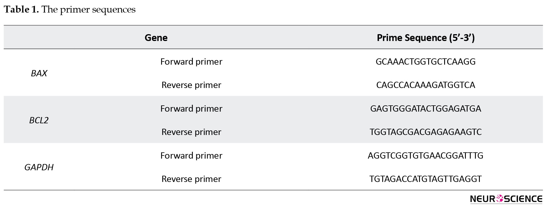
The entire cellular RNA was applied to prepare complementary DNA (cDNA) and used in the measurement of BCL-2 and BAX mRNA expression. Pars Tous Kit (Iran) was utilized to extract total RNA. The method and kit, as instructed by the manufacturer of Yekta Tajhiz Azma Kit (Iran), were used for cDNA preparation. Eventually, the cDNA was maintained at -20 °C. The housekeeping gene GADPH was used for normalization of target gene expression. The applied primes are shown in Table 1.
First, hippocampus samples were prepared, and then the purification of extracted RNA was performed. The high-quality RNAs were kept at -80 °C until they were used for cDNA synthesis. One milligram of RNA was converted to cDNA using the quantitect reverse transcription kit (Qiagen). To carry out real-time PCR, the primers were designed and underwent a large-scale search using the BLAST tool. Real-time PCR was conducted by executing the following cycling conditions: 95 °C for 10 min, and 40 cycles at 95 °C for 15 s, and 60 °C for 1 min. Every single complete amplification stage subsequently had a dissociation stage: At 95 for 15 s, 60 for 30 s, afterwards the temperature was boosted from 60 to 95 (at the rate of 0.03/s). Melting curve analysis was accomplished according to the dissociation stage data and reactions.
Cavalieri’s volumetric analysis
Cavalieri’s principle was used to assess the volume of the CA1 region. It is essential to ensure that all sections of the object of interest are parallel. The distance between them is known, and the first coronal section indiscriminately hits the object of interest. To estimate the cross-sections, coronal brain sections were stained with hematoxylin and eosin (H&E).
The total volume (V, mm3) of the CA1 region was calculated by Cavalier’s method, where V=ΣP (total number of the volume profiles counted in each rat’s hippocampus) × a/p (the area associated with each point) × t (the distance between the sampled sections).
Nissl staining
Sections of 10 micron thickness were air-dried after being placed directly onto gelatin-coated glass slides. The slides were dehydrated and cover-slipped with Entellan after being dyed with 1.0% cresyl violet. Each animal had eight photomicrographs taken (between the level of 2.3 and 5 mm posterior to bregma, according to the Paxinos atlas). A blinded investigator randomly picked three of them with a minimum distance of 40 microns and counted them using a light microscope at 400x magnification. Only pyramidal cells with visible nuclei and nucleoli were included in the study. Images were collected with a microscope (Olympus AX-70, Japan) at 400x magnification and analyzed with Image-Pro-Plus software, version 6.0 (LEICA DMLB, Germany).
Statistical analysis
Data are reported as Mean±SD. Analysis of variance (ANOVA) and least significant difference (LSD) methods are conducted for the comparison of different groups. In addition, a P<0.05 was identified as significant.
3. Results
Investigation of particle size of nanoemulsions
The shape of the graphs is a single peak, and the particle size distribution is narrow, indicating that the particles are small and uniform. The particle size is between 300 and 350 nanometers. Another device was used to ensure the data was accurate. The results of the sample size of black seed hydroalcoholic nano-extract are shown in Figure 1.
Transient global ischemia in humans occurs due to cardiac arrest or cardiac surgery, which causes selective and delayed nerve death, especially pyramidal neurons in the hippocampus (Tanaka et al., 2000). The key structure of the memory system is the hippocampus, which is used to study phenomena related to ischemia. However, specific hippocampal subsets illustrate different sensitivities to ischemia/hypoxia. The most sensitive area of the hippocampus is CA1. Even if it is exposed to a short-term ischemic attack, the mechanisms of delayed neuronal death leading to a significant reduction of nerve cells occur in this structure (Jiao et al., 2011). It has been reported that CA2-3 segments are relatively resistant to ischemia. Long-term ischemia due to ischemic recurrence causes changes in CA2-3 neurons, but the reduction of neurons, which is evident in this part, is rarely shown (Kirino & Sano, 1984). Nerve death due to global ischemia occurs up to 2-4 days after ischemia in rats, and the fetus is not recognizable. The role of apoptotic death and necrosis in ischemia-induced nerve cell death remains controversial (Yamashima & Oikawa, 2009; Graham & Chen, 2001).
However, the underlying mechanisms of ischemic death are still unclear. The molecular events that determine the fate of neurons could be investigated due to the significant delay between attack and the onset of death. It has been shown that extracts of herbs preserve protection against ischemic damage to various organs such as the kidneys, heart, and brain (Peng et al., 2003). Apoptosis, or programmed cell death, plays a critical role in neurodegenerative conditions and ischemia-induced brain damage. Among the key regulators of apoptosis are the BCL-2 family proteins, which include both pro-apoptotic and anti-apoptotic members. BAX (Bcl-2-associated X protein) promotes apoptosis by facilitating mitochondrial outer membrane permeabilization, whereas BCL-2 (B-cell lymphoma 2) inhibits this process and promotes cell survival. The ratio of BAX to BCL-2 is often used as an indicator of a cell’s susceptibility to apoptosis. Changes in the expression of BAX and BCL-2 genes can indicate whether a treatment exerts pro-survival or pro-apoptotic effects on neurons.
Therefore, assessing these markers helps evaluate the neuroprotective potential of experimental treatments (Antonsson & Martinou, 2000). In the past few years, scientists have worked to identify the active compounds in herbal medications and to understand how they work (Meddah et al., 2009). Black seed, or Nigella sativa, a member of the Ranunculaceae family, is one of these herbal treatments that is used for a broad range of diseases, including bronchial asthma, headache, allergies, gastrointestinal problems, hypertension, obesity, and various types of cancer. N. sativa seeds can also help reduce depression and have immunostimulatory effects on various inflammatory and immunologic diseases, including arthritis, colitis, and experimental allergic encephalomyelitis, as well as in sensitized animals, asthma patients, and chemical warfare victims, due to its documented component (Kaplan & Miller, 2000). Recently, clinical and animal studies on therapeutic drugs of black seed extracts have revealed that these drugs include a variety of effects such as anti inflammation (Hajhashemi et al., 2004) and modulation of the immune system (Tekeoglu., 2007), antioxidant activities (Kanter et al., 2006), and protection from light (Kanter et al., 2006). Recently, considerable attention has been focused on plants containing natural products and agents that can mitigate the damage caused by free radicals. The current research aims to explore the effect of hydroalcoholic and nano-hydroalcoholic extracts containing N. sativa on neurons in the CA1 area of the hippocampus in an ischemia model of the Wistar rats.
2. Materials and Methods
Study animals
Test subjects were 32 male Wistar rats, aged 8 weeks and weighing 200-250 g, which were accommodated under standard conditions (23±2 °C, 12/12 h light–dark cycle, relative humidity of 50±6%). They were provided with food and water in suitable amounts. All employed experimental procedures, such as caring and handling of the animals, were executed in compliance with the Tehran Medical Sciences Branch, Islamic Azad University Ethics Committee acts.
Study design
The test subjects were haphazardly divided into four groups of 8.
In the control group, the rats were not given any treatment or surgery.
In the ischemia group, after anesthesia, the carotid arteries of the rats were clamped for 20 minutes, followed by reperfusion.
In the experimental group 1, after ischemia and reperfusion for 20 minutes, the intraperitoneal injection of hydroalcoholic extract of black seed at a dose of 400 mg/kg was performed for 14 days straight.
In the experimental group 2, after ischemia and reperfusion for 20 minutes, the intraperitoneal injection of nano-hydroalcoholic extract of black seed at a dose of 400 mg/kg was performed for 14 consecutive days.
Twenty-four hours after the last injection, all animals underwent evaluation using the Y-maze behavioral test. Subsequently, the rat brains were removed for histological studies and gene evaluation.
Surgical process
After anesthesia by injection of pentobarbital sodium (40 mg/kg, IP), a vertical incision was made in the anterior region of the animal’s neck, and by pushing the sternocleidomastoid muscle, the common carotid arteries were exposed on both sides. After detachment of the vagus nerve, a microsurgical clamp was applied for 20 minutes to close the arteries. Then the clamps were removed and circulation was restored. During the surgery, the animal’s temperature was measured regularly using a thermometer, and a heat lamp stabilized the temperature at 37±0.5 °C. The cut was sutured with 04 silicon yarn. All the animals were monitored in separate cages until their conditions were stabilized (Dellu., 1992).
Y-maze behavioral test
A Y-maze apparatus was used to test short-term spatial memory. In simple words, a Y-maze is made up of three arms that cross each other at a 120° angle. It is preferred that test respondents examine the fresh arms with a higher frequency than a previously inspected arm. It was thought that revisiting a previously researched arm was a mistake. A lower tendency to explore the most recently visited arm indicated a better memory performance. The occurrence of entries into all three different arms (A, B, and C) in sequence was considered a valid alternation, reflecting genuine memory-based exploration. The total number of entries was recorded (Longa et al., 1989).
Preparation method of the hydroalcoholic extract of N. sativa
The alcoholic extraction of N. sativa was carried out using the maceration method, where the crushed grain was poured into the decanter’s funnel. Each time, 250 g of Nigella powder with 60% alcohol was added as the solvent. Then, the solution was placed at room temperature for 8-10 hours. Afterwards, the valve of the decanter was opened and the solution was passed through the filter twice. After drying the extract, it was measured, and 32% w/w extract was obtained (Tavakkoli et al., 2017).
Preparation method of nano-hydroalcoholic extract of N. sativa
First, the hydroalcoholic extract was poured into a container. Then, the alcohol was removed, and the extract was dried. After that, 10 g of the dried extract was dissolved in 100 mL of distilled water. The acquired hydroalcoholic extract was put into the ultrasonic probe for 1 h. Then,
The dynamic light scattering (DLS) was used to evaluate the nanoemulsion containing N. sativa. DLS is a physical method that is applied to actuate the distribution of particles in solutions and suspensions. Hydrodynamic diameter was used to measure the zeta potential, which serves as a marker to investigate the surface charge of the nanoemulsion. Additionally, transmission electron microscopy (TEM) was employed to examine the shape and size of the nanoemulsion (Tavakkoli et al., 2017).
Real-time PCR
The entire cellular RNA was applied to prepare complementary DNA (cDNA) and used in the measurement of BCL-2 and BAX mRNA expression. Pars Tous Kit (Iran) was utilized to extract total RNA. The method and kit, as instructed by the manufacturer of Yekta Tajhiz Azma Kit (Iran), were used for cDNA preparation. Eventually, the cDNA was maintained at -20 °C. The housekeeping gene GADPH was used for normalization of target gene expression. The applied primes are shown in Table 1.
First, hippocampus samples were prepared, and then the purification of extracted RNA was performed. The high-quality RNAs were kept at -80 °C until they were used for cDNA synthesis. One milligram of RNA was converted to cDNA using the quantitect reverse transcription kit (Qiagen). To carry out real-time PCR, the primers were designed and underwent a large-scale search using the BLAST tool. Real-time PCR was conducted by executing the following cycling conditions: 95 °C for 10 min, and 40 cycles at 95 °C for 15 s, and 60 °C for 1 min. Every single complete amplification stage subsequently had a dissociation stage: At 95 for 15 s, 60 for 30 s, afterwards the temperature was boosted from 60 to 95 (at the rate of 0.03/s). Melting curve analysis was accomplished according to the dissociation stage data and reactions.
Cavalieri’s volumetric analysis
Cavalieri’s principle was used to assess the volume of the CA1 region. It is essential to ensure that all sections of the object of interest are parallel. The distance between them is known, and the first coronal section indiscriminately hits the object of interest. To estimate the cross-sections, coronal brain sections were stained with hematoxylin and eosin (H&E).
The total volume (V, mm3) of the CA1 region was calculated by Cavalier’s method, where V=ΣP (total number of the volume profiles counted in each rat’s hippocampus) × a/p (the area associated with each point) × t (the distance between the sampled sections).
Nissl staining
Sections of 10 micron thickness were air-dried after being placed directly onto gelatin-coated glass slides. The slides were dehydrated and cover-slipped with Entellan after being dyed with 1.0% cresyl violet. Each animal had eight photomicrographs taken (between the level of 2.3 and 5 mm posterior to bregma, according to the Paxinos atlas). A blinded investigator randomly picked three of them with a minimum distance of 40 microns and counted them using a light microscope at 400x magnification. Only pyramidal cells with visible nuclei and nucleoli were included in the study. Images were collected with a microscope (Olympus AX-70, Japan) at 400x magnification and analyzed with Image-Pro-Plus software, version 6.0 (LEICA DMLB, Germany).
Statistical analysis
Data are reported as Mean±SD. Analysis of variance (ANOVA) and least significant difference (LSD) methods are conducted for the comparison of different groups. In addition, a P<0.05 was identified as significant.
3. Results
Investigation of particle size of nanoemulsions
The shape of the graphs is a single peak, and the particle size distribution is narrow, indicating that the particles are small and uniform. The particle size is between 300 and 350 nanometers. Another device was used to ensure the data was accurate. The results of the sample size of black seed hydroalcoholic nano-extract are shown in Figure 1.
DLS analysis
After the Nigella hydroalcoholic sample was prepared, DLS was employed to analyze particle size and distribution, and particle dispersion was examined using diagrams. The dispersion was between 294.4 and 357.1 nm, and a single peak diagram was obtained. The diameter of this diagram increases intensively when the size of particles exceeds 90 nm. If there are smaller particles with lower concentrations in the suspensions, they sometimes fade from the sensors, resulting in no intensity being reported. Following the stability test in 4 different environments, with one test conducted after one month and another after three months, the results remained consistent with the initial findings.
Investigation of particle size by zeta potential
The results of the zeta potential of N. sativa nano-hydroalcoholic extract are as follows (Figures 2 and 3).
Zeta potential analysis
Most fluids contain cations and anions, which are atoms with positive and negative charges. When the loaded particles are suspended in a liquid, the opposing load ions are absorbed into the particulate matter. It means that samples with negative loads attract positive ions from the fluid, and conversely, samples with a positive charge attract negative ions from the fluid. The ions near the surface of the particle are highly absorbed while the farther ions will have a loose bond, called the intrusion layer. Zeta potential ranges from -10 to +10, indicating the stability of the sample (Figure 4).
Behavioral test results
The results of the Y-maze test revealed that ischemia remarkably decreased the kinetic ability of test subjects. The aggregate of suitable feedback in the I/R group considerably declined in comparison with the control group. Furthermore, the longer the time of ischemia occurred, the worse the viciousness of neurological function was. Compared with the I/R group, the nano-hydroalcoholic extract of black seed recipient group showed significantly increased memory functions after I/R injury (Figures 4 and 5).
Results of the weight of rats in different groups
As a result of measuring the weight of rats before and after induction of ischemia and drug injection, it was observed that after surgery and during 14 days, when the experimental groups 1 and 2 were receiving the drug, one-way ANOVA test and LSD test showed a significant difference between ischemia group and experimental groups 1 and 2 (Figure 6).
Results of real-time PCR
The relative expression of BAX in the hippocampus of the I/R group was considerably higher than that of the control group, whereas the relative expression of BCL-2 mRNA was substantially lower. (Figures 7 and 8).
As shown in Figures 7 and 8, the treatment of rats with hydroalcoholic extract of black seed and nano-extract of hydroalcoholic black seed effectively decreased the relative expression of pro-apoptotic mRNAs BAX (Figure 7, P<0.05). It increased the relative expression of Bcl-2 in these groups (Figure 8, P<0.05).
Nissl staining results
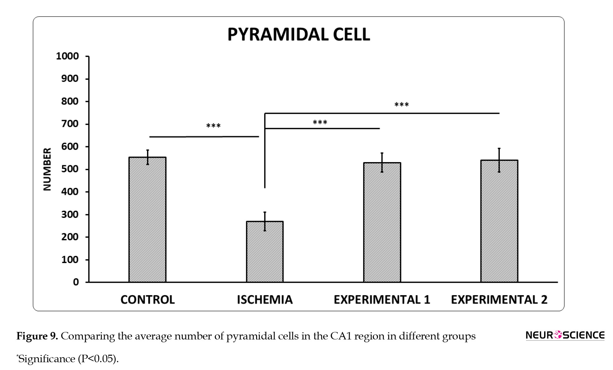
Nissl staining was used to determine viable pyramidal cells in the CA1 area. Cells with round, bright nuclei with an euchromatin appearance and multiple nuclei were considered as viable cells. Cells with dense, multifaceted, and heterochromatin nuclei were considered degenerative cells. The results of ANOVA showed that transient global I/R significantly decreased the number of pyramidal neurons (P<0.001). While the injection of hydroalcoholic extract and nano-hydroalcoholic extract containing N. sativa reversed the effect of transient global I/R on the number of neurons (P<0.05) (Figures 9 and 10).
Nissl staining results
Nissl staining was used to determine viable pyramidal cells in the CA1 area. Cells with round, bright nuclei with an euchromatin appearance and multiple nuclei were considered as viable cells. Cells with dense, multifaceted, and heterochromatin nuclei were considered degenerative cells. The results of ANOVA showed that transient global I/R significantly decreased the number of pyramidal neurons (P<0.001). While the injection of hydroalcoholic extract and nano-hydroalcoholic extract containing N. sativa reversed the effect of transient global I/R on the number of neurons (P<0.05) (Figures 9 and 10).
Most of the pyramidal cells were healthy. Note that a denotes healthy pyramidal cells, and b refers to degenerated pyramidal cells.
Results of stereological evaluation of the volume of the CA1 region of the hippocampus
The results of calculating the volume of the hippocampus CA1 region of 6 histological slides with H&E staining for each rat and a total of 6 rats in each group were observed. The volume in the ischemia group decreased significantly compared to the control group. Furthermore, the volume of the hippocampus CA1 area by injection of hydroalcoholic extract increased significantly compared to the injection of N. sativa nano-hydroalcoholic extract (Figure 11).
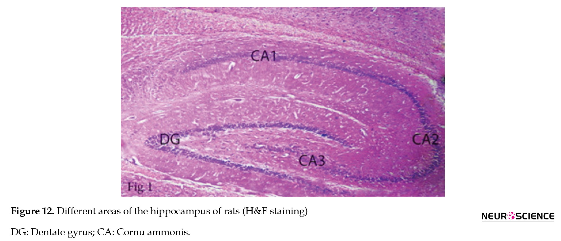
H&E staining were used to measure the volume of the CA1 region (Figure 12).
4. Discussion
Our findings showed that cerebral ischemia/reperfusion for 20 minutes caused delayed death of pyramidal cells in the CA1 region.
Ischemia-reperfusion injury is a pathological condition that has both local and systemic effects. The key element during ischemia is cell energy depletion, and the interplay of oxidative and microcirculatory stress, as well as inflammation and apoptosis, takes place during reperfusion (Soares et al., 2019).
Following ischemia, hippocampus may be affected by programmed death of cells. Oxidative stress caused by reactive oxygen species (ROS) production contributes to the programmed death of cells (Itō, 1984). Increased ROS levels inside the cell can lead to DNA and intracellular protein damage through oxidation, resulting in cell death.
In response to global cerebral ischemia, CA1 neurons in the hippocampus are particularly susceptible and experience selective, delayed degeneration. These pyramidal neurons play a crucial role in spatial learning and memory, and their degeneration causes learning and memory problems (Xu et al., 2021; Zuo et al., 2015).
Antiinflammatory and antioxidant medications should be investigated to provide additional treatment options that can help prevent or reduce these injuries, as current drugs have limited efficacy and offer little to no benefit to patients (Mucha et al., 2021). Antioxidants are compounds that can significantly slow down the oxidative reaction (Molitoris & Wagner, 1996). So far, many natural and synthetic antioxidants have been introduced for the treatment or prevention of patients associated with free radicals (Farahmand & Tajali, 2016). Medicinal plants always have an instrumental role in the management of global health care and are considered the best treatment for diseases (Dehpour et al., 2011). N. sativa is a medicinal plant that is highly consumed. In recent years, further attention has been paid to the medicinal and biological properties of N. sativa.
One of N. sativa’s possible qualities is the ability of one or more of its components to minimize tissue damage following ischemic/reperfusion injury due to antioxidant activity (Bayrak et al., 2008; Erşahin et al., 2011).
It has been reported that administration of antioxidant agents before local brain ischemia in laboratory studies protects against ischemic brain lesion (Lipton., 1999). Based on a review of the literature, thymoquinone, which is the most important extract of N. sativa, has many pharmacological properties, mostly related to its antioxidant properties. In this regard, Hosseinzadeh et al. (2007) reported that the administration of Nigella oils in the global ischemic model of the brain has reduced the amount of lipid peroxidation. Their findings can support the role of antioxidant properties in the protective effects of N. sativa in cerebral ischemia. Ziyyat et al. (1997) investigated the neuroprotective effects of thymoquinone on transient cerebral ischemia on the hippocampus in rats. Thymoquinone reduces malondialdehyde levels, increases reduced glutathione, catalase, and superoxide dismutase activity until it reaches its normal level. Thymoquinone is a promising factor in healing the damages caused by the destruction of neural cells (neurodegeneration), such as cerebral ischemia. N. sativa also enhances the activity of antioxidant enzymes such as superoxide dismutase and glutathione peroxidase, and with its antioxidant properties, reduces lipid peroxidation in biological membranes (Butt & Sultan, 2010). It is known that thymoquinone and its active metabolite thymohydroquinone can prevent lipid peroxidation by sweeping superoxide, radical hydroxyl, and single molecular oxygen (Kanter, 2008). In addition, thymoquinone can inhibit arachidonic acid metabolism and restrict ischemic brain damage caused by inflammation of cyclooxygenase and lipoxygenase pathways (El Mezayen et al., 2006).
Nanoparticle-based drug delivery systems have demonstrated higher potential in treating ischemic stroke and potentially other neurological disorders. Joachim et al. showed that the gelatin of osteopontin nanoparticles in a rat ischemic stroke model augments nerve protection in the nose (Joachim et al., 2014). Nagai et al. showed that intravenous administration of silvestazole nanoparticles improves acute ischemic damage in the brain following the ischemic/reperfusion (Nagai et al. 2015). According to the previous studies and the results of the current research concerning the function of N. sativa plant and its role in preventing the death of neural cells, it can be said that the hydroalcoholic extract and nano-hydroalcoholic extract of N. sativa can exert its protective effect on ischemia/reperfusion brain damages, which can be attributed to the properties of oxygen free radicalization of this plant. Our results showed a significant decrease in BAX expression and an increase in BCL-2 expression following treatment with [compound/extract], suggesting a shift toward anti-apoptotic signaling. This BAX/BCL-2 modulation indicates that the therapy may protect against ischemia-induced neuronal apoptosis by promoting cell survival pathways. Also, the N. sativa plant in nano form has more efficacy and higher adsorption than the hydroalcoholic extract. Still, since the role of other compounds in these plants, which has not been studied, and the role of other mechanisms, such as inhibition of inflammatory pathways, and also the possibility of a cumulative effect of these compounds, is not known, this issue needs further research and studies.
5. Conclusion
This research revealed that hydroalcoholic extract and nano-hydroalcoholic extract containing N. sativa, by preventing morphological changes and resulting cell death, have an effective role on neurons in the CA1 region of the hippocampus. Therefore, it seems that hydroalcoholic extract and nano-hydroalcoholic extract containing N. sativa through antiinflammatory and antioxidant mechanisms can be considered as a treatment candidate for ischemic brain damage.
Ethical Considerations
This study was approved by the Ethics Committee of Tehran Medical Sciences Branch, Islamic Azad University, Tehran, Iran (Code: IR.IAU.TMU.REC.1399.515). The ARRIVE ethical criteria were followed in all of the trials. During the research, the Guide for the Care and Use of Laboratory Animals (NIH Publication No. 86-23) was also followed.
Funding
This research did not receive any grant from funding agencies in the public, commercial, or non-profit sectors.
Authors' contributions
Conceptualization, supervision, data analysis, interpretation, review and editing: Zahra Nadia Sharifi and Atarodalsadat Mostavafinia; Methodology and experimental design: Mahsa Golmohammadi and Atarodalsadat Mostavafinia; Data collection and laboratory work: Mahsa Golmohammadi and Mohammad Mahdi Nazarnejad; Original draft preparation: Mohammad Mahdi Nazarnejad; Final approval: All authors.
Conflict of interest
The authors declared no conflict of interest.
Acknowledgments
The authors are grateful to the Herbal Pharmacology Research Center, Tehran Medical Sciences Branch, Islamic Azad University, Tehran, Iran, and all of the personnel For their direction and support,
References
Antonsson, B., & Martinou, J. C. (2000). The Bcl-2 protein family. Experimental Cell Research, 256(1), 50-57. [DOI:10.1006/excr.2000.4839] [PMID]
Bayrak, O., Bavbek, N., Karatas, O. F., Bayrak, R., Catal, F., & Cimentepe, E., et al. (2008). Nigella sativa protects against ischaemia/reperfusion injury in rat kidneys. Nephrology, Dialysis, Transplantation : Official Publication of the European Dialysis and Transplant Association - European Renal Association, 23(7), 2206–2212. [DOI:10.1093/ndt/gfm953] [PMID]
Butt, M. S., & Sultan, M. T. (2010). Nigella sativa: Reduces the risk of various maladies. Critical Reviews in Food Science and Nutrition, 50(7), 654-665. [DOI:10.1080/10408390902768797] [PMID]
Dehpour, A. A., Ebrahimzadeh, M. A., Roudgar, A., Nabavi, S.F., & Nabavi, S. M. (2011). Antioxidant and antibacterial activity of Consolida orientalis. World Acad Sci Eng Technol, 73:162-6. [Link]
Dellu, F., Mayo, W., Cherkaoui, J., Le Moal, M., & Simon, H. (1992). A two-trial memory task with automated recording: Study in young and aged rats. Brain Research, 588(1), 132-139. [DOI:10.1016/0006-8993(92)91352-F] [PMID]
Erşahin, M., Toklu, H. Z., Akakin, D., Yuksel, M., Yeğen, B. Ç., & Sener, G. (2011). The effects of Nigella sativa against oxidative injury in a rat model of subarachnoid hemorrhage. Acta Neurochirurgica, 153(2), 333-341. [DOI:10.1007/s00701-010-0853-9] [PMID]
El Mezayen, R., El Gazzar, M., Nicolls, M. R., Marecki, J. C., Dreskin, S. C., & Nomiyama, H. (2006). Effect of thymoquinone on cyclooxygenase expression and prostaglandin production in a mouse model of allergic airway inflammation. Immunology Letters, 106(1), 72-81. [DOI:10.1016/j.imlet.2006.04.012] [PMID]
Farahmand, M., & Tajali, M. (2016). [The effect of Salvia rhytidea extract on the Number of cells of different Layers of cerebellar cortex following ischemia reperfusion in rats (Persian)]. Armaghane Danesh, 21(6), 536-551. [Link]
Graham, S. H., & Chen, J. (2001). Programmed cell death in cerebral ischemia. Journal of Cerebral Blood Flow & Metabolism, 21(2), 99-109. [DOI:10.1097/00004647-200102000-00001] [PMID]
Hajhashemi, V., Ghannadi, A., & Jafarabadi, H. (2004). Black cumin seed essential oil, as a potent analgesic and antiinflammatory drug. Phytotherapy Research: An International Journal Devoted to Pharmacological and Toxicological Evaluation of Natural Product Derivatives, 18(3), 195-199. [DOI:10.1002/ptr.1390] [PMID]
Hosseinzadeh, H., Parvardeh, S., Asl, M. N., Sadeghnia, H. R., & Ziaee, T. (2007). Effect of thymoquinone and Nigella sativa seeds oil on lipid peroxidation level during global cerebral ischemia-reperfusion injury in rat hippocampus. Phytomedicine, 14(9), 621-627. [DOI:10.1016/j.phymed.2006.12.005] [PMID]
Itō, M. (1984). The cerebellum and neural control. New York: Raven Press. [Link]
Jiao, L., Zhang, J., Li, Z., Liu, H., Chen, Y., & Xu, S. (2011). Edaravone alleviates delayed neuronal death and long-dated cognitive dysfunction of hippocampus after transient focal ischemia in Wistar rat brains. Neuroscience, 182, 177-183. [DOI:10.1016/j.neuroscience.2011.01.017] [PMID]
Joachim, E., Kim, I. D., Jin, Y., Kim, K. K., Lee, J. K., & Choi, H. (2014). Gelatin nanoparticles enhance the neuroprotective effects of intranasally administered osteopontin in rat ischemic stroke model. Drug Delivery and Translational Research, 4(5-6), 395-399. [DOI:10.1007/s13346-014-0208-9] [PMID]
Kaplan, D. R., & Miller, F. D. (2000). Neurotrophin signal transduction in the nervous system. Current Opinion in Neurobiology, 10(3), 381-391. [DOI:10.1016/S0959-4388(00)00092-1] [PMID]
Kanter, M., Coskun, O., & Uysal, H. (2006). The antioxidative and antihistaminic effect of Nigella sativa and its major constituent, thymoquinone on ethanol-induced gastric mucosal damage. Archives of Toxicology, 80(4), 217-224. [DOI:10.1007/s00204-005-0037-1] [PMID]
Kanter, M., Coskun, O., Kalayci, M., Buyukbas, S., & Cagavi, F. (2006). Neuroprotective effects of Nigella sativa on experimental spinal cord injury in rats. Human & Experimental Toxicology, 25(3), 127-133. [DOI:10.1191/0960327106ht608oa] [PMID]
Kanter, M. (2008). Nigella sativa and derived thymoquinone prevents hippocampal neurodegeneration after chronic toluene exposure in rats. Neurochemical Research, 33(3), 579-588. [DOI:10.1007/s11064-007-9481-z] [PMID]
Kirino, T., & Sano, K. (1984). Selective vulnarablility in the gerbil hippocampus following transient ischemia. Actaneuropathologica, 62(3), 201-208. [DOI: 10.1007/BF00691853] [PMID]
Longa, E. Z., Weinstein, P. R., Carlson, S., & Cummins, R. (1989). Reversible middle cerebral artery occlusion without craniectomy in rats. Stroke, 20(1), 84-91. [DOI:10.1161/01.STR.20.1.84] [PMID]
Lipton, P. (1999). Ischemic cell death in brain neurons. Physiological Reviews, 79(4), 1431–1568. [DOI:10.1152/physrev.1999.79.4.1431] [PMID]
Meddah, B., Ducroc, R., Faouzi, M. E. A., Eto, B., Mahraoui, L., & Benhaddou-Andaloussi, A., et al. (2009). Nigella sativa inhibits intestinal glucose absorption and improves glucose tolerance in rats. Journal of Ethnopharmacology, 121(3), 419-424. [DOI:10.1016/j.jep.2008.10.040] [PMID]
Mucha, P., Skoczyńska, A., Małecka, M., Hikisz, P., & Budzisz, E. (2021). Overview of the Antioxidant and Anti-Inflammatory Activities of Selected Plant Compounds and Their Metal Ions Complexes. Molecules (Basel, Switzerland), 26(16), 4886. [DOI:10.3390/molecules26164886] [PMID]
Molitoris, B. A., & Wagner, M. C. (1996). Surface membrane polarity of proximal tubular cells: alterations as a basis for malfunction. Kidney International, 49(6), 1592-1597. [DOI:10.1038/ki.1996.231] [PMID]
Nagai, N., Yoshioka, C., Ito, Y., Funakami, Y., Nishikawa, H., & Kawabata, A. (2015). Intravenous administration of cilostazol nanoparticles ameliorates acute ischemic stroke in a cerebral ischemia/reperfusion-induced injury model. International Journal of Molecular Sciences, 16(12), 29329-29344. [DOI:10.3390/ijms161226166] [PMID]
Peng, H., Li, Y. F., & Sun, S. G. (2003). Effects of Ginkgo biloba extract on acute cerebral ischemia in rats analyzed by magnetic resonance spectroscopy. Acta Pharmacologica Sinica, 24(5), 467-471. [PMID]
Soares, R. O., Losada, D. M., Jordani, M. C., Évora, P., & Castro-e-Silva, O. (2019). Ischemia/reperfusion injury revisited: an overview of the latest pharmacological strategies. International Journal of Molecular Sciences, 20(20), 5034. [DOI:10.3390/ijms20205034] [PMID]
Tanaka, H., Grooms, S. Y., Bennett, M. V., & Zukin, R. S. (2000). The AMPAR subunit GluR2: Still front and center-stage. Brain Research, 886(1-2), 190-207. [DOI:10.1016/S0006-8993(00)02951-6]
Tavakkoli, A., Ahmadi, A., Razavi, B. M., & Hosseinzadeh, H. (2017). Black seed (Nigella sativa) and its constituent thymoquinone as an antidote or a protective agent against natural or chemical toxicities. Iranian Journal of Pharmaceutical Research: IJPR, 16(Suppl), 2-23. [PMID]
Tekeoglu, I., Dogan, A., Ediz, L., Budancamanak, M., & Demirel, A. (2007). Effects of thymoquinone (volatile oil of black cumin) on rheumatoid arthritis in rat models. Phytotherapy Research: An International Journal Devoted to Pharmacological and Toxicological Evaluation of Natural Product Derivatives, 21(9), 895-897. [DOI:10.1002/ptr.2143] [PMID]
Xu, H., Wang, E., Chen, F., Xiao, J., & Wang, M. (2021). Neuroprotective phytochemicals in experimental ischemic stroke: mechanisms and potential clinical applications. Oxidative Medicine and Cellular Longevity, 2021, 6687386. [DOI:10.1155/2021/6687386] [PMID]
Yamashima, T., & Oikawa, S. (2009). The role of lysosomal rupture in neuronal death. Progress in Neurobiology, 89(4), 343-358. [DOI:10.1016/j.pneurobio.2009.09.003] [PMID]
Zuo, W., Zhang, W., Han, N., & Chen, N. H. (2015). Compound IMM-H004, a novel coumarin derivative, protects against CA1 cell loss and spatial learning impairments resulting from transient global ischemia. CNS Neuroscience & Therapeutics, 21(3), 280–288. [DOI:10.1111/cns.12364] [PMID]
Ziyyat, A., Legssyer, A., Mekhfi, H., Dassouli, A., Serhrouchni, M., & Benjelloun, W. (1997). Phytotherapy of hypertension and diabetes in oriental Morocco. Journal of Ethnopharmacology, 58(1), 45-54. [DOI:10.1016/S0378-8741(97)00077-9] [PMID]
Type of Study: Original |
Subject:
Cellular and molecular Neuroscience
Received: 2025/03/6 | Accepted: 2025/06/10 | Published: 2025/07/1
Received: 2025/03/6 | Accepted: 2025/06/10 | Published: 2025/07/1
Send email to the article author
| Rights and permissions | |
 |
This work is licensed under a Creative Commons Attribution-NonCommercial 4.0 International License. |

Copyright © The Author(s);
This is an open access article distributed under the terms of the Creative Commons Attribution License (CC-By-NC), which permits use, distribution, and reproduction in any medium, provided the original work is properly cited and is not used for commercial purposes.
Contact Information