
Volume 16, Issue 4 (July & August 2025)
BCN 2025, 16(4): 805-818 |
Back to browse issues page



Download citation:
BibTeX | RIS | EndNote | Medlars | ProCite | Reference Manager | RefWorks
Send citation to:


BibTeX | RIS | EndNote | Medlars | ProCite | Reference Manager | RefWorks
Send citation to:
Kazmi S, Yazdanfar N, Seyedaghamiri F, Pishgahi A, Yousefi M, Ghadiri T, et al . Intranasal Autologous Conditioned Serum Attenuates Memory Impairment After mPFC Ischemia in Mice. BCN 2025; 16 (4) :805-818
URL: http://bcn.iums.ac.ir/article-1-3158-en.html
URL: http://bcn.iums.ac.ir/article-1-3158-en.html
Sareh Kazmi1 

 , Neda Yazdanfar1 , Fatemehsadat Seyedaghamiri1 , Alireza Pishgahi2 , Mehdi Yousefi3 , Tahereh Ghadiri4 , Saeed Sadigh-Eteghad5 , Mehdi Farhoudi *6
, Neda Yazdanfar1 , Fatemehsadat Seyedaghamiri1 , Alireza Pishgahi2 , Mehdi Yousefi3 , Tahereh Ghadiri4 , Saeed Sadigh-Eteghad5 , Mehdi Farhoudi *6
1- Department of Neuroscience, Faculty of Advanced Medical Sciences, Tabriz University of Medical Sciences, Tabriz, Iran. & Neurosciences Research Center, Tabriz University of Medical Sciences, Tabriz, Iran.
2- Physical Medicine and Rehabilitation Specialist, Motahari General Hospital, Isfahan, Iran.
3- Immunology Research Center, Tabriz University of Medical Sciences, Tabriz, Iran.
4- Department of Neuroscience, Faculty of Advanced Medical Sciences, Tabriz University of Medical Sciences, Tabriz, Iran.
5- Neurosciences Research Center, Tabriz University of Medical Sciences, Tabriz, Iran.
6- Neurosciences Research Center, Aging Research Institute, Tabriz University of Medical Sciences, Tabriz, Iran.
2- Physical Medicine and Rehabilitation Specialist, Motahari General Hospital, Isfahan, Iran.
3- Immunology Research Center, Tabriz University of Medical Sciences, Tabriz, Iran.
4- Department of Neuroscience, Faculty of Advanced Medical Sciences, Tabriz University of Medical Sciences, Tabriz, Iran.
5- Neurosciences Research Center, Tabriz University of Medical Sciences, Tabriz, Iran.
6- Neurosciences Research Center, Aging Research Institute, Tabriz University of Medical Sciences, Tabriz, Iran.
Keywords: Ischemic stroke, Autologous conditioned serum (ACS), Phosphorylated tau (p-tau), Interleukin-1 receptor antagonist (IL-1RA), Insulin-like growth factor-1 (IGF-I)
Full-Text [PDF 2234 kb]
| Abstract (HTML)
ACS effect on social memory in the social interaction test
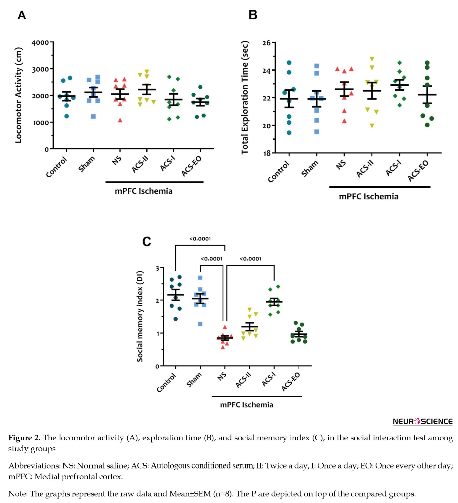
There was no significant difference in locomotor activity, as assessed by the total distance traveled and total exploration time (Figures 2A and 2B).
ACS increases IGF-1 and IL-1RA levels after mPFC ischemia
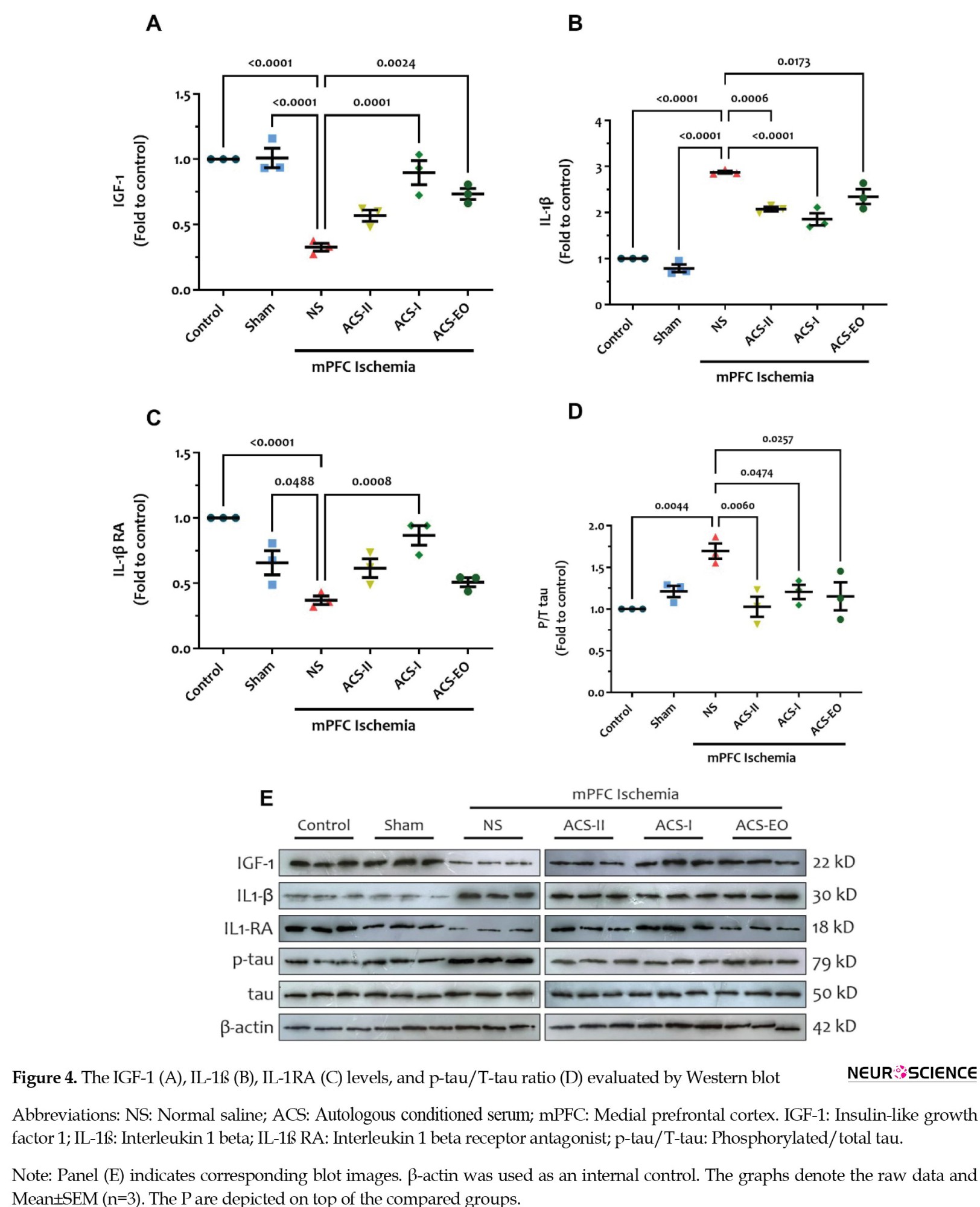
Quantitative analysis revealed significant reductions in both IGF-1 and IL-1RA levels in the NS-treated ischemic group compared to the control and sham groups (P<0.05). The ACS-I and ACS-EO groups exhibited an increase in IGF-1 expression; however, an increase in IL-1RA levels was observed only in the ACS-I group (at least P<0.01) (Figures 4A and 4C).
4. Discussion
According to the results of the present study, intranasal administration of ACS improves spatial and social memory impairments in mPFC ischemia in mice. The observed therapeutic effects likely result from dual mechanisms: Upregulation of neuroprotective IGF-1 and downregulation of pathological tau phosphorylation (p-tau/t-tau ratio) in peri-infarct areas. Additionally, ACS reduced pro-inflammatory cytokine (IL-1β) and augmented the level of IL-1RA cytokine in the ischemic area.
The global burden of stroke and dementia is rising at an alarming rate, with current epidemiological projections estimating that by 2050, there will be approximately 200 million stroke survivors and 106 million individuals living with dementia worldwide. This dramatic increase, representing a 150% surge in stroke prevalence and near-tripling of dementia cases compared to 2020 baselines, reflects the compounding effects of aging populations, particularly in low- and middle-income countries, alongside insufficient management of vascular risk factors and limited therapeutic breakthroughs for neurodegenerative processes (Brainin et al., 2020). Accordingly, it has been reported that approximately one in every three or four stroke survivors develops some form of cognitive decline or dementia (Kalaria et al., 2016), which hinders successful recovery. Clinical and imaging data have identified that functional outcomes following a stroke depend on factors such as the type of stroke, its severity, location, number of occurrences, and the volume of insult (Onyike, 2006; Rost et al., 2022). In this context, no significant differences have been reported in cognitive assessment between stroke subtypes (Aam et al., 2020); however, greater survival and incidence rates of ischemic stroke compared to hemorrhagic stroke leave a higher number of ischemic stroke survivors experiencing cognitive decline (Perna & Temple, 2015). Additionally, the anatomical location of the lesion is considered an influential factor for cognitive decline and is a strategic contributor to developing future dementia in stroke patients (Munsch et al., 2016; Zhao et al., 2017).
The PFC primarily contributes to a wide range of cognitive and executive functions, which are governed by lateral, medial, and orbitofrontal subregions exerting top-down control over other cortical and subcortical domains (Jobson et al., 2021). Data from electrophysiological and behavioral analyses suggest that the mPFC is implicated in high cognitive functions, including spatial working memory, attention, social behavior, and decision making (Carboni et al., 2024; Giacometti Giordani et al., 2023; Wirt & Hyman, 2017). Studies utilizing experimental models have indicated impaired recognition memory, spatial memory, cognitive inflexibility, and anxiety-related behavior following mPFC ischemia (Happ et al., 2020; Livingston-Thomas et al., 2015). Results from the current investigation revealed that mPFC ischemia increased latency and time spent in the error zone in the Lashley III maze, indicating impaired spatial learning. Similarly, it has been documented that focal ischemia in mPFC decreased displacement index in the what-where-which test, demonstrating impaired episodic memory (Sadigh-Eteghad et al., 2018). Moreover, the mPFC inactivation exhibited impaired inhibition of retrieval of non-practiced items, which is an essential phenomenon for successful memory recall by blocking retrieval of inappropriate competing interfaces (Wu et al., 2014). Scott et al. demonstrated that the inactivation of the mPFC led to a decline in odor working memory, and disconnecting its circuit with the mediodorsal thalamus resulted in dysfunction in exploratory motor activity (Scott et al., 2020).
We also found that mPFC photothrombotic ischemic induction dramatically declines the social memory index in the social interaction test. The mPFC has been proposed as a crucial foundation for social memory and social novelty encoding, facilitated by its extensive projections to limbic structures and top-down control over various circuits (Ko, 2017). The pharmacological blockade of the mPFC has been shown to block social recognition memory (Marcondes et al., 2020). Moreover, the long-term excitatory-inhibitory imbalance of hippocampal projections to mPFC has been found to impair social memory without affecting other social interactions, suggesting that any disruptions in the neural substrates of this circuit interfere with social memory processing (Phillips et al., 2019). It has been demonstrated that mPFC parvalbumin neurons mediate social behavior by targeting the hippocampal-dependent social memory pathway (Sun et al., 2020).
The IGF-1 is a trophic signal promoting maturation and growth in most tissues. Data from clinical studies have proven the positive role of IGF-1 overexpression in human motor and cognitive performances, while its suppression is observed in individuals with AD (Ferreira, 2021). Downregulation of IGF-1 compromised its capacity to promote the polarization of M2 microglia, suppressed microglia reactive oxygen species, and M1 phenotype markers like TNF-α, and inhibited astrocytic response to stimuli (Labandeira-Garcia et al., 2017). Furthermore, systemic inflammation has been shown to increase levels of the inflammatory cytokine TNF-α and IGF binding protein-1 (IGFBP-1), while reducing IGF-1 levels by approximately half in affected tissues, such as the brain (Fan et al., 1995). Additionally, intravenous IGF-1 improves the cognitive and neurological functions of rats subjected to ischemic stroke. This improvement was associated with the restoration of IGF-1 levels in the hippocampus, cortex, and amygdala of the affected rats. This exogenously increased levels of IGF-1 led to decreased levels of IL-6, IL-1β, and TNF-α in the plasma and hippocampus, as well as down-regulation of phospho-tau protein in the hippocampus and reduced cortical infarct volume (Yang et al., 2020).
P-tau is considered a shared biomarker between stroke and neurodegenerative disease. The higher plasma level of p-tau in patients with a history of stroke, regardless of whether they have dementia or not, compared to control individuals (Tang et al., 2018), could be related to the blood-brain barrier disruption following a stroke (Kurzepa et al., 2010). Subcortical focal ischemia was proposed to upregulate the microtubule-associated regulatory kinase, leading to microtubule destabilization by phosphorylation of tau at serine-262 in the microtubule-binding domain (Hayden et al., 2019). Tau phosphorylation at this site is believed to act as a gateway phosphorylation site to enhance further phosphorylation and tau aggregation (Biernat et al., 1993). Neuroinflammation as a pathological hallmark of ischemic stroke develops within minutes and continues for days and weeks following the attack (Anrather & Iadecola, 2016). Cells in the ischemic core and peri-infarct zone begin to release inflammatory molecules such as cytokines and chemokines into the systemic circulation, leading to the infiltration of peripheral immune cells into the brain, which then activate the host immune system (Wu et al., 2022; Xu et al., 2020). The microglia express pro- and anti-inflammatory cytokines in infarct and peri-infarct lesion sites (Lambertsen et al., 2019; Lambertsen et al., 2019).
IL-1RA endogenously inhibits IL-1α and IL-1β, working by blocking the IL-1β signaling pathway to reduce inflammation (Harrell et al., 2020). Due to the low concentration of IL-1RA in affected tissues, boosting the level of IL-1RA could be an appealing target to achieve maximum therapeutic benefits (Camargo Garbin & Morris, 2021). Recently, immunotherapy in clinical stroke trials has gained attention for targeting innate and adaptive responses by lowering microglia activation, suppressing immune cell migration to the brain, and modulating the IL-1β signaling pathway (Drieu et al., 2018). ACS is a bioproduct enriched in IL-1RA, IL-4, IL-10, and growth factors (Shakouri et al., 2021), which has been clinically used in orthopedic disorders and is currently in clinical trials for COVID-19 (Fotouhi et al., 2018; Shakouri et al., 2020). In the present investigation, we demonstrated that intranasal ACS therapy buffered spatial memory and social memory dysfunctions, which probably occurred through its anti-inflammatory abilities to upregulate IGF-1 and IL-1RA and downregulate IL-1β and p-tau in the mPFC of animals that underwent photothrombotic ischemic stroke. Inhibiting IL-1RA has been claimed to increase vascular inflammation by increasing IL-1 signaling, introducing it as an anti-inflammatory therapeutic option in conditions such as ischemic stroke. In this context, it has been reported that blocking IL-1α after stroke reduced endothelial activation and expression of adhesion molecules. This condition led to a decrease in penumbral mononuclear phagocyte content and neurotoxic mediators like matrix metalloprotease, ultimately modulating cerebral injury (Liberale et al., 2021). In agreement, data from a phase II placebo-controlled clinical trial in ischemic and hemorrhagic stroke cases has shown that the subcutaneous injection of recombinant human IL-1RA reduced the plasma level of IL-6 and C-reactive protein and also decreased the extent of edema observed in CT scans of hemorrhagic stroke patients (Parry-Jones et al., 2023; Smith et al., 2018). It is suggested that IL-1RA enhanced the effectiveness of tissue plasminogen activator (tPA) treatment, a gold standard therapy for ischemic stroke, by inhibiting IL-1 to elevate the levels of endogenous tPA inhibitor-1 (Smith et al., 2018). It has been accepted that microglia IL-1β increases neuronal tau phosphorylation through p38-MAPk signaling pathway, thereby disrupt cytoskeleton assembly and axon stabilization leading to neuronal cell death (Li et al., 2003). Indeed, IL-1β serves as a vital substrate for tauopathy conditions’ pathological biomarkers (Maphis et al., 2015). IL-1β-treated microglial culture has been shown to upregulate the level of secreted fragment of the β-amyloid precursor protein (s-APP). Given the positive role of pre-treatment with conditioned medium from sAPP-activated microglia with IL-RA in down-regulation of of β- p-tau, α-synuclein, and p38-MAPk, IL-1R could be assigned as a promising therapy in tauopathy conditions (Griffin et al., 2006).
5. Conclusion
The data from our study showed that chronic ACS administration after stroke significantly improved spatial and social interaction memories. Additionally, ACS therapy notably increased the protein abundance of IGF-1 and IL-1RA, while simultaneously decreasing the production of IL-1β and p-tau in the mPFC. However, to fully elucidate the cellular and molecular mechanisms, further exact studies are needed to examine how ACS mediates the observed neuroprotective effects against brain ischemia.
Ethical Considerations
Compliance with ethical guidelines
This study was approved by the Ethics Committee of Tabriz University of Medical Sciences, Tabriz, Iran (Code: IR.TBZMED.VCR.REC.1399.403) and performed following the guidelines of the National Institutes of Health (NIH) (Publication No. 85–23, revised 1985).
Funding
This research was supported by a grant from the Tabriz University of Medical sciences, Tabriz, Iran (Grant No.: 60948).
Authors' contributions
Conceptualization: Sareh Kazmi, Alireza Pishgahi, Mehdi Yousefi, Tahereh Ghadiri, Saeed Sadigh-Eteghad, and Saeed Sadigh-Eteghad; Investigation: Sareh Kazmi, Fatemehsadat Seyedaghamiri, Alireza Pishgahi and Mehdi Yousefi: Data curation: Sareh Kazmi; Formal analysis: Sareh Kazmi and Saeed Sadigh-Eteghad; Validation: Saeed Sadigh-Eteghad; Writing: Sareh Kazmi, Neda Yazdanfar, Tahereh Ghadiri, Saeed Sadigh-Eteghad, and Mehdi Farhoudi; final approval: All authors.
Conflict of interest
The authors declared no conflict of interest.
Acknowledgments
The authors would like to appreciate the Tabriz University of Medical sciences for supporting of this work.
References
Aam, S., Einstad, M. S., Munthe-Kaas, R., Lydersen, S., Ihle-Hansen, H., & Knapskog, A. B., et al. (2020). Post-stroke cognitive impairment-impact of follow-up time and stroke subtype on severity and cognitive profile: The Nor-COAST study. Frontiers in Neurology, 11, 699. [DOI:10.3389/fneur.2020.00699] [PMID]
Angadi, D. S., Macdonald, H., & Atwal, N. (2020). Autologous cell-free serum preparations in the management of knee osteoarthritis: What is the current clinical evidence? Knee Surgery & Related Research, 32(1), 16. [DOI:10.1186/s43019-020-00036-5] [PMID]
Anrather, J., & Iadecola, C. (2016). Inflammation and stroke: An overview. Neurotherapeutics, 13(4), 661-670. [DOI:10.1007/s13311-016-0483-x] [PMID]
Bhasin, A., Srivastava, M., Bhatia, R., Mohanty, S., Kumaran, S., & Bose, S. (2012). Autologous intravenous mononuclear stem cell therapy in chronic ischemic stroke. Journal of Stem Cells & Regenerative Medicine, 8(3), 181-189. [DOI:10.46582/jsrm.0803011] [PMID]
Bi, M., Gladbach, A., van Eersel, J., Ittner, A., Przybyla, M., & van Hummel, A., et al. (2017). Tau exacerbates excitotoxic brain damage in an animal model of stroke. Nature Communications, 8(1), 473. [DOI:10.1038/s41467-017-00618-0] [PMID]
Biernat, J., Gustke, N., Drewes, G., Mandelkow, E. M., & Mandelkow, E. (1993). Phosphorylation of Ser262 strongly reduces binding of tau to microtubules: distinction between PHF-like immunoreactivity and microtubule binding. Neuron, 11(1), 153–163. [DOI:10.1016/0896-6273(93)90279-z] [PMID]
Brainin, M., Feigin, V. L., Norrving, B., Martins, S. C. O., Hankey, G. J., & Hachinski, V., et al. (2020). Global prevention of stroke and dementia: The WSO Declaration. The Lancet Neurology, 19(6), 487-488. [DOI:10.1016/S1474-4422(20)30141-1] [PMID]
Brassai, A., Suvanjeiev, R. G., Bán, E. G., & Lakatos, M. (2015). Role of synaptic and nonsynaptic glutamate receptors in ischaemia induced neurotoxicity. Brain Research Bulletin, 112, 1-6. [DOI:10.1016/j.brainresbull.2014.12.007] [PMID]
Camargo Garbin, L., & Morris, M. J. (2021). A comparative review of autologous conditioned serum and autologous protein solution for treatment of osteoarthritis in horses. Frontiers in Veterinary Science, 8, 602978. [DOI:10.3389/fvets.2021.602978] [PMID]
Carboni, E., Ibba, M., Carboni, E., & Carta, A. R. (2024). Adolescent stress differentially modifies dopamine and norepinephrine release in the medial prefrontal cortex of adult rats. Progress in Neuro-Psychopharmacology and Biological Psychiatry, 134, 111055. [DOI:10.1016/j.pnpbp.2024.111055] [PMID]
Chen, J., Li, Y., Wang, L., Zhang, Z., Lu, D., & Lu, M., et al. (2001). Therapeutic benefit of intravenous administration of bone marrow stromal cells after cerebral ischemia in rats. Stroke, 32(4), 1005-1011. [DOI:10.1161/01.str.32.4.1005] [PMID]
Chen, J., Sanberg, P. R., Li, Y., Wang, L., Lu, M., & Willing, A. E., et al. (2001). Intravenous administration of human umbilical cord blood reduces behavioral deficits after stroke in rats. Stroke, 32(11), 2682–2688. [DOI:10.1161/hs1101.098367] [PMID]
Chen, X., & Jiang, H. (2019). Tau as a potential therapeutic target for ischemic stroke. Aging, 11(24), 12827–12843. [DOI:10.18632/aging.102547] [PMID]
Doeppner, T. R., Herz, J., Görgens, A., Schlechter, J., Ludwig, A. K., & Radtke, S., et al. (2015). Extracellular vesicles improve post-stroke neuroregeneration and prevent postischemic immunosuppression. Stem Cells Translational Medicine, 4(10), 1131–1143. [DOI:10.5966/sctm.2015-0078] [PMID]
Drieu, A., Levard, D., Vivien, D., & Rubio, M. (2018). Anti-inflammatory treatments for stroke: From bench to bedside. Therapeutic Advances in Neurological Disorders, 11, 1756286418789854. [DOI:10.1177/1756286418789854] [PMID]
Fan, J., Li, Y. H., Bagby, G. J., & Lang, C. H. (1995). Modulation of inflammation-induced changes in insulin-like growth factor (IGF)-I and IGF binding protein-1 by anti-TNF antibody. Shock, 4(1), 21-26. [DOI:10.1097/00024382-199507000-00003] [PMID]
Ferreira, S. T. (2021). Brain insulin, insulin‐like growth factor 1 and glucagon‐like peptide 1 signalling in Alzheimer’s disease. Journal of Neuroendocrinology, 33(4), e12959. [DOI:10.1111/jne.12959] [PMID]
Fotouhi, A., Maleki, A., Dolati, S., Aghebati-Maleki, A., & Aghebati-Maleki, L. (2018). Platelet rich plasma, stromal vascular fraction and autologous conditioned serum in treatment of knee osteoarthritis. Biomedicine & Pharmacotherapy, 104, 652–660. [DOI:10.1016/j.biopha.2018.05.019] [PMID]
Frizziero, A., Giannotti, E., Oliva, F., Masiero, S., & Maffulli, N. (2013). Autologous conditioned serum for the treatment of osteoarthritis and other possible applications in musculoskeletal disorders. British Medical Bulletin, 105, 169–184. [DOI:10.1093/bmb/lds016] [PMID]
Giacometti Giordani, L., Crisafulli, A., Cantarella, G., Avenanti, A., & Ciaramelli, E. (2023). The role of posterior parietal cortex and medial prefrontal cortex in distraction and mind-wandering. Neuropsychologia, 188, 108639. [PMID]
Gong, P., Zou, Y., Zhang, W., Tian, Q., Han, S., & Xu, Z., et al. (2021). The neuroprotective effects of insulin-like growth factor 1 via the Hippo/YAP signaling pathway are mediated by the PI3K/AKT cascade following cerebral ischemia/reperfusion injury. Brain Research Bulletin, 177, 373-387. [DOI:10.1016/j.brainresbull.2021.10.017] [PMID]
Griffin, W. S., Liu, L., Li, Y., Mrak, R. E., & Barger, S. W. (2006). Interleukin-1 mediates Alzheimer and Lewy body pathologies. Journal of Neuroinflammation, 3, 5. [DOI:10.1186/1742-2094-3-5] [PMID]
Happ, D. F., Wegener, G., & Tasker, R. A. (2020). Effect of ischemic lesions in medial prefrontal cortex and nucleus accumbens on affective behavior in rats. Behavioural Brain Research, 378, 112234. [DOI:10.1016/j.bbr.2019.112234] [PMID]
Harrell, C. R., Markovic, B. S., Fellabaum, C., Arsenijevic, N., Djonov, V., & Volarevic, V. (2020). The role of Interleukin 1 receptor antagonist in mesenchymal stem cell-based tissue repair and regeneration. BioFactors (Oxford, England), 46(2), 263–275. [DOI:10.1002/biof.1587] [PMID]
Hayden, E. Y., Putman, J., Nunez, S., Shin, W. S., Oberoi, M., & Charreton, M., et al. (2019). Ischemic axonal injury up-regulates MARK4 in cortical neurons and primes tau phosphorylation and aggregation. Acta Neuropathologica Communications, 7(1), 135. [DOI:10.1186/s40478-019-0783-6] [PMID]
Houlton, J., Barwick, D., & Clarkson, A. N. (2021). Frontal cortex stroke-induced impairment in spatial working memory on the trial-unique nonmatching-to-location task in mice. Neurobiology of Learning and Memory, 177, 107355. [DOI:10.1016/j.nlm.2020.107355] [PMID]
Jobson, D. D., Hase, Y., Clarkson, A. N., & Kalaria, R. N. (2021). The role of the medial prefrontal cortex in cognition, ageing and dementia. Brain Communications, 3(3), fcab125.[DOI:10.1093/braincomms/fcab125] [PMID]
Kalaria, R. N., Akinyemi, R., & Ihara, M. (2016). Stroke injury, cognitive impairment and vascular dementia. Biochimica et Biophysica Acta, 1862(5), 915–925. [DOI:10.1016/j.bbadis.2016.01.015] [PMID]
Ko, J. (2017). Neuroanatomical substrates of rodent social behavior: The medial prefrontal cortex and its projection patterns. Frontiers in Neural Circuits, 11, 41. [DOI:10.3389/fncir.2017.00041] [PMID]
Kurzepa, J., Bielewicz, J., Grabarska, A., Stelmasiak, Z., Stryjecka-Zimmer, M., & Bartosik-Psujek, H. (2010). Matrix metalloproteinase-9 contributes to the increase of tau protein in serum during acute ischemic stroke. Journal of Clinical Neuroscience, 17(8), 997-999. [DOI:10.1016/j.jocn.2010.01.005] [PMID]
Labandeira-Garcia, J. L., Costa-Besada, M. A., Labandeira, C. M., Villar-Cheda, B., & Rodríguez-Perez, A. I. (2017). Insulin-like growth factor-1 and neuroinflammation. Frontiers in Aging Neuroscience, 9, 365. [DOI:10.3389/fnagi.2017.00365] [PMID]
Lambertsen, K. L., Finsen, B., & Clausen, B. H. (2019). Post-stroke inflammation-target or tool for therapy? Acta Neuropathologica, 137(5), 693-714. [DOI:10.1007/s00401-018-1930-z] [PMID]
Li, W., Ye, A., Ao, L., Zhou, L., Yan, Y., & Hu, Y., et al. (2020). Protective mechanism and treatment of neurogenesis in cerebral ischemia. Neurochemical Research, 45(10), 2258-2277. [DOI:10.1007/s11064-020-03092-1] [PMID]
Li, Y., Liu, L., Barger, S. W., & Griffin, W. S. (2003). Interleukin-1 mediates pathological effects of microglia on tau phosphorylation and on synaptophysin synthesis in cortical neurons through a p38-MAPK pathway. The Journal of Neuroscience: The Official Journal of the Society for Neuroscience, 23(5), 1605–1611. [DOI:10.1523/jneurosci.23-05-01605.2003] [PMID]
Liberale, L., Ministrini, S., Carbone, F., Camici, G. G., & Montecucco, F. (2021). Cytokines as therapeutic targets for cardio- and cerebrovascular diseases. Basic Research in Cardiology, 116(1), 23. [DOI:10.1007/s00395-021-00863-x] [PMID]
Livingston-Thomas, J. M., Jeffers, M. S., Nguemeni, C., Shoichet, M. S., Morshead, C. M., & Corbett, D. (2015). Assessing cognitive function following medial prefrontal stroke in the rat. Behavioural Brain Research, 294, 102–110. [DOI:10.1016/j.bbr.2015.07.053] [PMID]
Maphis, N., Xu, G., Kokiko-Cochran, O. N., Jiang, S., Cardona, A., & Ransohoff, R. M., et al. (2015). Reactive microglia drive tau pathology and contribute to the spreading of pathological tau in the brain. Brain, 138(Pt 6), 1738-1755. [DOI:10.1093/brain/awv081] [PMID]
Marcondes, L. A., Nachtigall, E. G., Zanluchi, A., de Carvalho Myskiw, J., Izquierdo, I., & Furini, C. R. G. (2020). Involvement of medial prefrontal cortex NMDA and AMPA/kainate glutamate receptors in social recognition memory consolidation. Neurobiology of Learning and Memory, 168, 107153. [DOI:10.1016/j.nlm.2019.107153] [PMID]
Munsch, F., Sagnier, S., Asselineau, J., Bigourdan, A., Guttmann, C. R., & Debruxelles, S., et al. (2016). Stroke location is an independent predictor of cognitive outcome. Stroke, 47(1), 66-73. [DOI:10.1161/STROKEAHA.115.011242] [PMID]
Onyike, C. U. (2006). Cerebrovascular disease and dementia. International Review of Psychiatry (Abingdon, England), 18(5), 423–431. [DOI:10.1080/09540260600935421] [PMID]
Parry-Jones, A. R., Stocking, K., MacLeod, M. J., Clarke, B., Werring, D. J., & Muir, K. W., et al. (2023). Phase II randomised, placebo-controlled, clinical trial of interleukin-1 receptor antagonist in intracerebral haemorrhage: BLOcking the cytokine IL-1 in ICH (BLOC-ICH). European Stroke Journal, 8(3), 819–827.[DOI:10.1177/23969873231185208] [PMID]
Perna, R., & Temple, J. (2015). Rehabilitation outcomes: Ischemic versus hemorrhagic strokes. Behavioural Neurology, 2015, 891651. [DOI:10.1155/2015/891651] [PMID]
Phillips, M. L., Robinson, H. A., & Pozzo-Miller, L. (2019). Ventral hippocampal projections to the medial prefrontal cortex regulate social memory. Elife, 8, e44182. [DOI:10.7554/eLife.44182] [PMID]
Pluta, R., Czuczwar, S. J., Januszewski, S., & Jabłoński, M. (2021). The many faces of post-ischemic tau protein in brain neurodegeneration of the Alzheimer’s disease type. Cells, 10(9), 2213. [DOI:10.3390/cells10092213] [PMID]
Pluta, R., Kiś, J., Januszewski, S., Jabłoński, M., & Czuczwar, S. J. (2022). Cross-talk between amyloid, tau protein and free radicals in post-ischemic brain neurodegeneration in the form of Alzheimer’s disease proteinopathy. Antioxidants, 11(1), 146. [DOI:10.3390/antiox11010146] [PMID]
Rost, N. S., Brodtmann, A., Pase, M. P., van Veluw, S. J., Biffi, A., & Duering, M., et al. (2022). Post-stroke cognitive impairment and dementia. Circulation Research, 130(8), 1252-1271. [DOI:10.1161/CIRCRESAHA.122.319951] [PMID]
Sadigh-Eteghad, S., Geranmayeh, M. H., Majdi, A., Salehpour, F., Mahmoudi, J., & Farhoudi, M. (2018). Intranasal cerebrolysin improves cognitive function and structural synaptic plasticity in photothrombotic mouse model of medial prefrontal cortex ischemia. Neuropeptides, 71, 61-69. [DOI:10.1016/j.npep.2018.07.002] [PMID]
Scott, G. A., Liu, M. C., Tahir, N. B., Zabder, N. K., Song, Y., & Greba, Q., et al. (2020). Roles of the medial prefrontal cortex, mediodorsal thalamus, and their combined circuit for performance of the odor span task in rats: Analysis of memory capacity and foraging behavior. Learning & Memory, 27(2), 67-77. [DOI:10.1101/lm.050195.119] [PMID]
Seyedaghamiri, F., Farajdokht, F., Vatandoust, S. M., Mahmoudi, J., Khabbaz, A., & Sadigh-Eteghad, S. (2021). Sericin modulates learning and memory behaviors by tuning of antioxidant, inflammatory, and apoptotic markers in the hippocampus of aged mice. Molecular Biology Reports, 48(2), 1371-1382. [DOI:10.1007/s11033-021-06195-2] [PMID]
Shakouri, S. K., Dolati, S., Santhakumar, J., Thakor, A. S., & Yarani, R. (2021). Autologous conditioned serum for degenerative diseases and prospects. Growth Factors, 39(1-6), 59-70. [DOI:10.1080/08977194.2021.2012467] [PMID]
Shakouri, S. K., Roshangar, L., & Mahmoodpoor, A. (2020). Intratracheal administration of autologus conditioned serum for COVID-19 associated respiratory distress syndrome. Journal of Critical Care, 60, 209–211. [DOI:10.1016/j.jcrc.2020.08.016] [PMID]
Smith, C. J., Hulme, S., Vail, A., Heal, C., Parry-Jones, A. R., & Scarth, S., et al. (2018). SCIL-STROKE (subcutaneous interleukin-1 receptor antagonist in ischemic stroke): A randomized controlled phase 2 trial. Stroke, 49(5), 1210-1216. [DOI:10.1161/STROKEAHA.118.020750] [PMID]
Sun, Q., Li, X., Li, A., Zhang, J., Ding, Z., & Gong, H., et al. (2020). Ventral hippocampal-prefrontal interaction affects social behavior via parvalbumin positive neurons in the medial prefrontal cortex. iScience, 23(3), 100894. [DOI:10.1016/j.isci.2020.100894] [PMID]
Tang, S. C., Yang, K. C., Chen, C. H., Yang, S. Y., Chiu, M. J., & Wu, C. C., et al. (2018). Plasma β-amyloids and tau proteins in patients with vascular cognitive impairment. Neuromolecular Medicine, 20(4), 498–503. [DOI:10.1007/s12017-018-8513-y] [PMID]
Uchihara, T., Nakamura, A., Arai, T., Ikeda, K., & Tsuchiya, K. (2004). Microglial tau undergoes phosphorylation-independent modification after ischemia. Glia, 45(2), 180-187. [DOI:10.1002/glia.10318] [PMID]
Wehling, P., Moser, C., Frisbie, D., Wayne McIlwraith, C., Kawcak, C. E., & Krauspe, R., et al. (2007). Autologous conditioned serum in the treatment of orthopedic diseases. BioDrugs Clinical Immunotherapeutics, Biopharmaceuticals and Gene Therapy, 21(5), 323–332. [DOI:10.2165/00063030-200721050-00004] [PMID]
Wirt, R. A., & Hyman, J. M. (2017). Integrating spatial working memory and remote memory: Interactions between the medial prefrontal cortex and hippocampus. Brain Sciences, 7(4), 43. [DOI:10.3390/brainsci7040043] [PMID]
Wu, F., Liu, Z., Zhou, L., Ye, D., Zhu, Y., & Huang, K., et al. (2022). Systemic immune responses after ischemic stroke: From the center to the periphery. Frontiers in Immunology, 13, 911661.[DOI:10.3389/fimmu.2022.911661] [PMID]
Wu, J. Q., Peters, G. J., Rittner, P., Cleland, T. A., & Smith, D. M. (2014). The hippocampus, medial prefrontal cortex, and selective memory retrieval: Evidence from a rodent model of the retrieval‐induced forgetting effect. Hippocampus, 24(9), 1070-1080. [DOI:10.1002/hipo.22291] [PMID]
Xu, S., Lu, J., Shao, A., Zhang, J. H., & Zhang, J. (2020). Glial cells: Role of the immune response in ischemic stroke. Frontiers in Immunology, 11, 294. [DOI:10.3389/fimmu.2020.00294] [PMID]
Yang, J., Wu, S., Hou, L., Zhu, D., Yin, S., & Yang, G., et al. (2020). Therapeutic effects of simultaneous delivery of nerve growth factor mRNA and protein via exosomes on cerebral ischemia. Molecular Therapy. Nucleic Acids, 21, 512–522. [DOI:10.1016/j.omtn.2020.06.013] [PMID]
Yang, W., Li, G., Cao, K., Ma, P., Guo, Y., & Tong, W., et al. (2020). Exogenous insulin-like growth factor 1 attenuates acute ischemic stroke-induced spatial memory impairment via modulating inflammatory response and tau phosphorylation. Neuropeptides, 83, 102082. [DOI:10.1016/j.npep.2020.102082] [PMID]
Zhao, L., Biesbroek, J. M., Shi, L., Liu, W., Kuijf, H. J., & Chu, W. W., et al. (2018). Strategic infarct location for post-stroke cognitive impairment: A multivariate lesion-symptom mapping study. Journal of Cerebral Blood Flow & Metabolism, 38(8), 1299-1311. [DOI:10.1177/0271678X17728162] [PMID]
Zhou, J., Du, T., Li, B., Rong, Y., Verkhratsky, A., & Peng, L. (2015). Crosstalk between MAPK/ERK and PI3K/AKT signal pathways during brain ischemia/reperfusion. ASN Neuro, 7(5), 1759091415602463. [DOI:10.1177/1759091415602463] [PMID]
Full-Text:
1. Introduction
The main neurological cause of death and disability worldwide is stroke, which has a narrow therapeutic window (Yang et al., 2020). Stroke is characterized by two foremost subtypes of blood flow disruption: Ischemic and hemorrhagic. Ischemic stroke, which accounts for 70%-85% of cases, is more common and often the focus of extensive research. However, there is an increasing awareness in societies about stroke prevention to protect against sensory, motor, and cognitive deficits caused by it (Li et al., 2020). The underlying mechanisms of cognitive impairments following an ischemic stroke have remained poorly understood, posing significant challenges in the daily lives of patients. These challenges include difficulties with language, executive function, visuospatial cognition, episodic memory, and working memory, highlighting the urgent need for further research (Houlton et al., 2021).
Of particular interest is why the prefrontal cortex (PFC)-induced ischemic model can be used in preclinical studies to enhance understanding of interventional research and assessment. The PFC plays a pivotal role in multidimensional, hierarchical, and top-down cognitive processing, and is also associated with normal age-related cognitive decline (Houlton et al., 2021). Given that ischemia in the PFC and frontal cortices leads to impaired reversal learning, spatial memory, cognitive flexibility, and coherence between the PFC and hippocampus (Jobson & Hase, 2021), we conducted a study using a focal medial PFC photothrombotic lesion in mice.
When ischemia occurs, it obstructs blood flow to the brain, leading to a decrease in oxygen supply and disruption of metabolic and internal balance. The activation of glutamate receptors mainly causes ischemic cell death due to changes in ion distribution and intracellular calcium concentration, which leads to excitotoxicity (Bi et al., 2017; Brassai et al., 2015). Brain damage in ischemia is driven by the overactivation of the N-methyl-D-aspartate (NMDA) receptors, which induces excitotoxicity. Tau, a protein highly expressed in neurons, regulates and stabilizes microtubules. The pathological transformation of tau in Alzheimer disease (AD) and frontotemporal dementia (FTD), characterized by hyperphosphorylation and tangle formation, results in functional deficits that contribute to disease progression (Uchihara et al., 2004). Following ischemia, hyperphosphorylated tau (p-tau) persists in neurons, similar to changes seen in AD. Research has shown that reducing p-tau levels can protect against excitotoxic deficits in the acute phase of stroke through ERK (extracellular signal-regulated kinase) signaling (Gong et al., 2021; Uchihara et al., 2004; Zhou et al., 2015). Inflammatory molecules severely disrupt normal tau functions, while misfolded tau, in turn, exacerbates inflammation, although this does not occur during the acute phase of stroke. During acute inflammation, microglia reduce the oligomerization of tau. P-tau accumulation can trigger apoptosis through endoplasmic reticulum-associated degradation. Conversely, inhibiting tau phosphorylation is crucial for protecting against ischemic conditions. In clinical settings, levels of tau in serum, plasma, or cerebrospinal fluid can be used to predict stroke outcomes and deficits (Chen & Jiang, 2019; Pluta et al., 2021; Pluta et al., 2022).
The term “autologous” refers to serum derived from syngeneic mice (same strain, age, and genetic background) rather than from the same individual mouse (Bhasin et al., 2012; Chen et al., 2001; Chen et al., 2001; Doeppner et al., 2015). The concept of utilizing cytokine inhibitors and growth factors for therapeutic purposes first emerged in the late 1970s to early 1980s. In orthopedic applications, growth factors have gained prominence for their capacity to modulate disease pathophysiology and promote functional tissue regeneration. Autologous conditioned serum (ACS) exemplifies this approach by delivering a concentrated cocktail of endogenous anti-inflammatory and anabolic factors to injured sites (Frizziero et al., 2013; Wehling et al., 2007). ACS is generated by incubating venous blood with medical-grade glass beads, a process that activates peripheral blood leukocytes to secrete anti-inflammatory mediators, particularly interleukin-1 receptor antagonist (IL-1RA). Following incubation, the serum is isolated via centrifugation and can be either stored for future applications or administered directly to the target organs. Since 1998, ACS, marketed as Orthokine, has been utilized in orthopedic patients and experimental animal models (Angadi et al., 2020).
2. Materials and Methods
Animals
Adult male C57BL/6 mice (10-12 weeks old, weighing approximately 25 g) were procured from the Animal Laboratory Center of the Pasteur Institute of Iran. The animals were kept in standard cages (8 animals per cage) under controlled conditions (24±2 °C) on a 12/12-hour light/dark cycle. They were provided with standard pellet food and tap water ad libitum. All experimental procedures were approved by the Ethics Committee of Tabriz University of Medical sciences and performed following the guidelines of the National Institutes of Health (NIH) Publication No. 85–23, revised 1985).
Preparation of ACS
For this purpose, we used the Heila kit (Roham Cell, Canada). Whole blood (300–500 μL per animal) was collected from the mice via intracardiac puncture into non-heparinized syringes containing medical-grade beads. Whole blood was maintained at 37 °C in a temperature-controlled incubator for 8 hours before centrifugation (4000×g, 10 min). The supernatant (ACS) was collected under sterile conditions and stored at −70 °C. The ACS was analyzed for IL-1Ra and insulin-like growth factor 1 (IGF-1) levels using the ELISA method according to the kit protocols.
Groups and administration
After one week of acclimatization to laboratory conditions, the animals were randomly divided into 6 groups (n=8 per group): Control, sham, mPFC ischemia + normal saline (NS), mPFC ischemia + ACS-II (received ACS intranasally twice daily at 12-hour intervals), mPFC ischemia + ACS-I (received ACS intranasally once daily), mPFC ischemia + ACS-EO (received ACS intranasally once every other day). The control group received NS injections without any surgical intervention, while the sham group underwent sham surgery followed by NS administration. The experimental groups were subjected to mPFC ischemia and subsequently treated with either NS or ACS. The animals in the NS-received groups were administered 48 μL of NS intranasally once a day. ACS was administered at a dosage of 48 μL per session for one week via the intranasal route. During administration, the ACS was alternated between the right and left nares at one-minute intervals.
Photothrombotic ischemia model induction
The bilateral photothrombotic mPFC ischemia model was established using the following protocol: Animals were anesthetized with isoflurane (5% for induction, 2% for maintenance) and secured in a stereotaxic apparatus. The skull overlying the mPFC (centered at 2.2 mm anterior to bregma) was surgically exposed and demarcated with sterile ink. Before illumination, Rose Bengal (150 μg/g body weight; Sigma-Aldrich, St. Louis, MO, USA) was administered intraperitoneally as a photosensitizing agent. Five minutes post-injection, the marked cortical region was irradiated for 10 minutes (continuous wave: 532 nm laser, 70 mW output power, 2 mm beam diameter) to induce localized thrombosis.
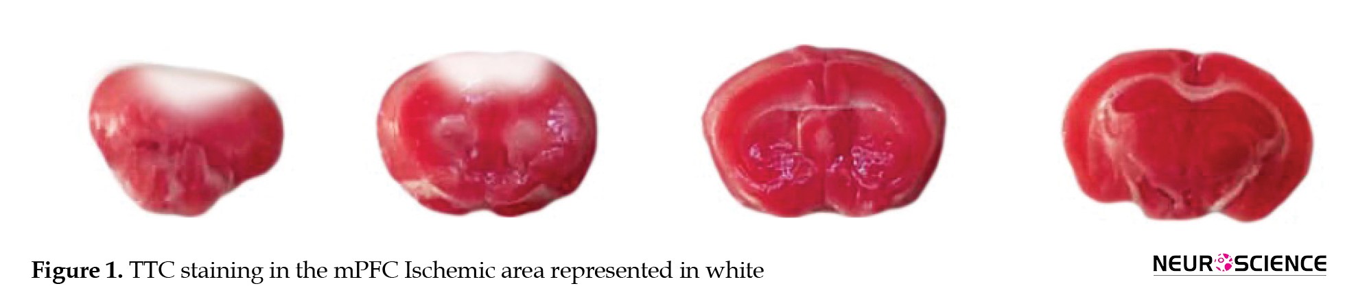
The surgical part was sutured after the procedure. Following photothrombotic induction, the animals were immediately transferred to a temperature-controlled recovery cage (maintained at 28-30 °C) and monitored until full ambulation returned. Postoperative analgesia (e.g. buprenorphine, 0.05 mg/kg) was administered subcutaneously every 8-12 hours for 24 hours. Animals were individually housed with free access to food and water during the study, with daily assessments of neurological function and wound healing. Ischemia induction was confirmed using TTC staining 48 hours after surgery. The same procedure was performed for the sham group animals, except they were not exposed to laser light. Their incisions were sutured as part of the protocol. This model is both safe and conservative, as no mortality was observed in any study group.
TTC staining for cerebral infarct assessment
2,3,5-Triphenyltetrazolium chloride (TTC) staining was performed to evaluate ischemic damage according to established protocols. Forty-eight hours post-ischemia, mice were euthanized under deep anesthesia (ketamine/xylazine: 90/10 mg/kg, IP) and transcardially perfused with ice-cold PBS. Brains were rapidly extracted and sectioned into 3-mm coronal slices using a brain matrix.
The sections were incubated in 2% TTC solution (w/v in PBS, pH 7.4) at 37 °C for 20 minutes, protected from light, then fixed in 4% paraformaldehyde. Viable tissue-stained brick-red due to mitochondrial dehydrogenase activity, while infarcted regions remained unstained (white).
Behavioral tests
Social interaction test
This test was performed to assess social memory. A square Plexiglas® box (57×45×30 cm), consisting of three interconnected compartments separated by two plexiglass walls, was used for the assessment. Each outer compartment contains a wire cage. The test consisted of three stages: Habituation, sociability assessment, and social memory evaluation, each lasting 10 minutes. At the first step, the mouse was placed in the central compartment and allowed to explore all three chambers freely. In the next step, a stranger mouse of the same age, sex, and weight was placed in one wire cage, while the other wire cage remained empty. The test mouse was then allowed to explore all three compartments freely. At the final step, the first stranger mouse remained in one wire cage, and a second stranger mouse of the same age and weight was placed in the opposite wire cage. The test mouse was again allowed to explore all compartments freely. Interaction time was scored when the subject mouse sniffed within 2 cm of the cages. Throughout all three stages, the animals’ behaviors and movements were recorded using a camera mounted above the apparatus (Seyedaghamiri et al., 2021).
Lashley maze test
This study used the Lashley III maze test to examine spatial memory. The maze consisted of three parts: A start box, a labyrinth, and a bonus box, all made of plexiglass. The arms of the maze had four parallel lines. Food was withheld from the animals 8 hours before the test to increase motivation. Some food was placed in the bonus box as a reward.
At the beginning of the test, the animal was located in the start box and permitted to move freely through the arms of the maze for 6 minutes to find the reward box. Once the animal found the reward, the test was terminated. This test was done for 5 consecutive days. During all stages of the test, the movement of the animals was recorded by a camera mounted above the maze. The latency to reach the bonus box and the number of errors (frequency of entries into incorrect arms) were recorded as parameters of interest.
Behavioral assessment methodology
Behavioral parameters were quantified using EthoVision™ XT video tracking system (v15.0, Noldus Information Technology, Wageningen, Netherlands). All testing apparatus was acquired from Arman Poshtiban Teb Co. (Tabriz, Iran) and standardized before experimentation.
Between trials, apparatus surfaces were thoroughly cleaned with 70% ethanol to eliminate residual olfactory cues, followed by a 5-minute drying period to ensure complete ethanol evaporation.
Tissue collection and processing
After behavioral testing, animals were perfused with PBS under ketamine/xylazine anesthesia (90/10 mg/kg). Brains were either dissected and frozen at -70 °C for immunoblotting or fixed in 4% Paraformaldehyde (PFA) for histology.
Western blot analysis of inflammatory and tau-related proteins
To evaluate the protein expression levels of IL-1β, IL-1RA, total tau (t-tau), phosphorylated tau (p-tau), and IGF-1, we performed Western blot analysis according to standardized protocols. Brain tissue samples were homogenized in RIPA lysis buffer (50 mM Tris-HCl, pH 7.4, 150 mM NaCl, 1% NP-40, 0.5% sodium deoxycholate, 0.1% sodium dodecyl sulfate [SDS]) supplemented with protease and phosphatase inhibitor cocktail. The homogenates were centrifuged at 14000×g for 20 min at 4 °C to remove insoluble debris. Protein concentrations were determined using the Bradford protein assay with bovine serum albumin as a standard.
Protein samples (20 μg per lane) were mixed with 2x Laemmli sample buffer containing 4% SDS and 10% 2-mercaptoethanol, boiled for 5 min, and separated by SDS-PAGE on 12% polyacrylamide gels. The separated proteins were then electrophoretically transferred onto 0.2-μm pore size polyvinylidene difluoride membranes (Bio-Rad Laboratories, Hercules, CA, USA) using a wet transfer system at 100 V for 90 min at 4 °C.
Membranes were blocked with 5% bovine serum albumin (BSA; Sigma-Aldrich, St. Louis, MO, USA) in Tris-buffered saline containing 0.1% Tween-20 (TBST) for 75 min at room temperature. The membranes were then incubated overnight at 4 °C with the following primary antibodies diluted in blocking buffer: rabbit anti-tau (1:1000; ab76128, Abcam), rabbit anti-phospho-tau (1:1000; ab92676, Abcam), rabbit anti-IGF-1 (1:800; ab9572, Abcam), rabbit anti-IL-1RA (1:1000; ab175392, Abcam), rabbit anti-IL-1β (1:800; ab254360, Abcam), and mouse anti-β-actin (1:5000; ab8227, Abcam).
After three washes with TBST (10 min each), membranes were incubated for 1 h at room temperature with horseradish peroxidase (HRP)-conjugated goat anti-rabbit IgG (1:5000; ab6721, Abcam) or goat anti-mouse IgG (1:5000; ab6789, Abcam) secondary antibodies. Protein bands were visualized using enhanced chemiluminescence substrate and imaged using a chemiluminescence detection system. Band intensities were quantified by densitometric analysis using ImageJ software (National Institutes of Health, Bethesda, MD, USA), with β-actin serving as the loading control for normalization.
Histological assessment of infarct volume by hematoxylin and eosin (H&E) staining
The extent of cerebral infarction was evaluated using standard H&E staining. Following fixation in 4% paraformaldehyde, the brain tissues were processed through a graded ethanol series (70%, 80%, 90%, and 100%) for dehydration, cleared in xylene, and embedded in paraffin blocks. Using a rotary microtome (DS-8402, Daeshin Precision, Korea), 12 consecutive (serial) 5-μm thick coronal sections were obtained from the medial PFC (mPFC) region, spanning the entire ischemic lesion. The sections were mounted on poly-L-lysine-coated slides and stained using Mayer’s hematoxylin solution for 8 minutes, followed by eosin Y counterstaining for 1 minute. After dehydration through an ascending alcohol series and xylene clearing, sections were coverslipped with Entellan mounting medium (Merck, Germany).
The infarct volume was quantified using image analysis software (Image4j version 0.7, NIH, Bethesda, MD, USA) based on the Equation 1:
1. Infarct area×Number of sections×Distance between sections.
Statistical analysis
All statistical analyses were conducted using GraphPad Prism software, version 8.01 (GraphPad Software Inc., La Jolla, CA, USA). For comparisons among multiple groups, we employed one-way analysis of variance (ANOVA) or 2-way ANOVA. Following significant ANOVA results (P<0.05), we performed Tukey’s honest significant difference (HSD) post-hoc tests for all pairwise comparisons. Results are presented as Mean±SEM throughout the manuscript, with individual data points shown in all graphs to demonstrate data distribution.
3. Results
ACS composition
The predominant cytokines in ACS were IL-1RA and IGF-I. The IL-1RA concentration in ACS was 10224.3±498.6 pg/mL, compared to 200.3±1.2 pg/mL in the unconditioned serum. This difference represents an approximately 50-fold increase in IL-1RA levels. Similarly, the IGF-I concentration in ACS was 119.2±0.44 pg/mL, compared to 22.2±0.41 pg/mL in the unconditioned serum, indicating a roughly 5-fold increase.
Model approval using TTC staining
TTC staining confirmed the induction of ischemia in the target cortical area (Figure 1) 48 hours after model induction.
The main neurological cause of death and disability worldwide is stroke, which has a narrow therapeutic window (Yang et al., 2020). Stroke is characterized by two foremost subtypes of blood flow disruption: Ischemic and hemorrhagic. Ischemic stroke, which accounts for 70%-85% of cases, is more common and often the focus of extensive research. However, there is an increasing awareness in societies about stroke prevention to protect against sensory, motor, and cognitive deficits caused by it (Li et al., 2020). The underlying mechanisms of cognitive impairments following an ischemic stroke have remained poorly understood, posing significant challenges in the daily lives of patients. These challenges include difficulties with language, executive function, visuospatial cognition, episodic memory, and working memory, highlighting the urgent need for further research (Houlton et al., 2021).
Of particular interest is why the prefrontal cortex (PFC)-induced ischemic model can be used in preclinical studies to enhance understanding of interventional research and assessment. The PFC plays a pivotal role in multidimensional, hierarchical, and top-down cognitive processing, and is also associated with normal age-related cognitive decline (Houlton et al., 2021). Given that ischemia in the PFC and frontal cortices leads to impaired reversal learning, spatial memory, cognitive flexibility, and coherence between the PFC and hippocampus (Jobson & Hase, 2021), we conducted a study using a focal medial PFC photothrombotic lesion in mice.
When ischemia occurs, it obstructs blood flow to the brain, leading to a decrease in oxygen supply and disruption of metabolic and internal balance. The activation of glutamate receptors mainly causes ischemic cell death due to changes in ion distribution and intracellular calcium concentration, which leads to excitotoxicity (Bi et al., 2017; Brassai et al., 2015). Brain damage in ischemia is driven by the overactivation of the N-methyl-D-aspartate (NMDA) receptors, which induces excitotoxicity. Tau, a protein highly expressed in neurons, regulates and stabilizes microtubules. The pathological transformation of tau in Alzheimer disease (AD) and frontotemporal dementia (FTD), characterized by hyperphosphorylation and tangle formation, results in functional deficits that contribute to disease progression (Uchihara et al., 2004). Following ischemia, hyperphosphorylated tau (p-tau) persists in neurons, similar to changes seen in AD. Research has shown that reducing p-tau levels can protect against excitotoxic deficits in the acute phase of stroke through ERK (extracellular signal-regulated kinase) signaling (Gong et al., 2021; Uchihara et al., 2004; Zhou et al., 2015). Inflammatory molecules severely disrupt normal tau functions, while misfolded tau, in turn, exacerbates inflammation, although this does not occur during the acute phase of stroke. During acute inflammation, microglia reduce the oligomerization of tau. P-tau accumulation can trigger apoptosis through endoplasmic reticulum-associated degradation. Conversely, inhibiting tau phosphorylation is crucial for protecting against ischemic conditions. In clinical settings, levels of tau in serum, plasma, or cerebrospinal fluid can be used to predict stroke outcomes and deficits (Chen & Jiang, 2019; Pluta et al., 2021; Pluta et al., 2022).
The term “autologous” refers to serum derived from syngeneic mice (same strain, age, and genetic background) rather than from the same individual mouse (Bhasin et al., 2012; Chen et al., 2001; Chen et al., 2001; Doeppner et al., 2015). The concept of utilizing cytokine inhibitors and growth factors for therapeutic purposes first emerged in the late 1970s to early 1980s. In orthopedic applications, growth factors have gained prominence for their capacity to modulate disease pathophysiology and promote functional tissue regeneration. Autologous conditioned serum (ACS) exemplifies this approach by delivering a concentrated cocktail of endogenous anti-inflammatory and anabolic factors to injured sites (Frizziero et al., 2013; Wehling et al., 2007). ACS is generated by incubating venous blood with medical-grade glass beads, a process that activates peripheral blood leukocytes to secrete anti-inflammatory mediators, particularly interleukin-1 receptor antagonist (IL-1RA). Following incubation, the serum is isolated via centrifugation and can be either stored for future applications or administered directly to the target organs. Since 1998, ACS, marketed as Orthokine, has been utilized in orthopedic patients and experimental animal models (Angadi et al., 2020).
2. Materials and Methods
Animals
Adult male C57BL/6 mice (10-12 weeks old, weighing approximately 25 g) were procured from the Animal Laboratory Center of the Pasteur Institute of Iran. The animals were kept in standard cages (8 animals per cage) under controlled conditions (24±2 °C) on a 12/12-hour light/dark cycle. They were provided with standard pellet food and tap water ad libitum. All experimental procedures were approved by the Ethics Committee of Tabriz University of Medical sciences and performed following the guidelines of the National Institutes of Health (NIH) Publication No. 85–23, revised 1985).
Preparation of ACS
For this purpose, we used the Heila kit (Roham Cell, Canada). Whole blood (300–500 μL per animal) was collected from the mice via intracardiac puncture into non-heparinized syringes containing medical-grade beads. Whole blood was maintained at 37 °C in a temperature-controlled incubator for 8 hours before centrifugation (4000×g, 10 min). The supernatant (ACS) was collected under sterile conditions and stored at −70 °C. The ACS was analyzed for IL-1Ra and insulin-like growth factor 1 (IGF-1) levels using the ELISA method according to the kit protocols.
Groups and administration
After one week of acclimatization to laboratory conditions, the animals were randomly divided into 6 groups (n=8 per group): Control, sham, mPFC ischemia + normal saline (NS), mPFC ischemia + ACS-II (received ACS intranasally twice daily at 12-hour intervals), mPFC ischemia + ACS-I (received ACS intranasally once daily), mPFC ischemia + ACS-EO (received ACS intranasally once every other day). The control group received NS injections without any surgical intervention, while the sham group underwent sham surgery followed by NS administration. The experimental groups were subjected to mPFC ischemia and subsequently treated with either NS or ACS. The animals in the NS-received groups were administered 48 μL of NS intranasally once a day. ACS was administered at a dosage of 48 μL per session for one week via the intranasal route. During administration, the ACS was alternated between the right and left nares at one-minute intervals.
Photothrombotic ischemia model induction
The bilateral photothrombotic mPFC ischemia model was established using the following protocol: Animals were anesthetized with isoflurane (5% for induction, 2% for maintenance) and secured in a stereotaxic apparatus. The skull overlying the mPFC (centered at 2.2 mm anterior to bregma) was surgically exposed and demarcated with sterile ink. Before illumination, Rose Bengal (150 μg/g body weight; Sigma-Aldrich, St. Louis, MO, USA) was administered intraperitoneally as a photosensitizing agent. Five minutes post-injection, the marked cortical region was irradiated for 10 minutes (continuous wave: 532 nm laser, 70 mW output power, 2 mm beam diameter) to induce localized thrombosis.
The surgical part was sutured after the procedure. Following photothrombotic induction, the animals were immediately transferred to a temperature-controlled recovery cage (maintained at 28-30 °C) and monitored until full ambulation returned. Postoperative analgesia (e.g. buprenorphine, 0.05 mg/kg) was administered subcutaneously every 8-12 hours for 24 hours. Animals were individually housed with free access to food and water during the study, with daily assessments of neurological function and wound healing. Ischemia induction was confirmed using TTC staining 48 hours after surgery. The same procedure was performed for the sham group animals, except they were not exposed to laser light. Their incisions were sutured as part of the protocol. This model is both safe and conservative, as no mortality was observed in any study group.
TTC staining for cerebral infarct assessment
2,3,5-Triphenyltetrazolium chloride (TTC) staining was performed to evaluate ischemic damage according to established protocols. Forty-eight hours post-ischemia, mice were euthanized under deep anesthesia (ketamine/xylazine: 90/10 mg/kg, IP) and transcardially perfused with ice-cold PBS. Brains were rapidly extracted and sectioned into 3-mm coronal slices using a brain matrix.
The sections were incubated in 2% TTC solution (w/v in PBS, pH 7.4) at 37 °C for 20 minutes, protected from light, then fixed in 4% paraformaldehyde. Viable tissue-stained brick-red due to mitochondrial dehydrogenase activity, while infarcted regions remained unstained (white).
Behavioral tests
Social interaction test
This test was performed to assess social memory. A square Plexiglas® box (57×45×30 cm), consisting of three interconnected compartments separated by two plexiglass walls, was used for the assessment. Each outer compartment contains a wire cage. The test consisted of three stages: Habituation, sociability assessment, and social memory evaluation, each lasting 10 minutes. At the first step, the mouse was placed in the central compartment and allowed to explore all three chambers freely. In the next step, a stranger mouse of the same age, sex, and weight was placed in one wire cage, while the other wire cage remained empty. The test mouse was then allowed to explore all three compartments freely. At the final step, the first stranger mouse remained in one wire cage, and a second stranger mouse of the same age and weight was placed in the opposite wire cage. The test mouse was again allowed to explore all compartments freely. Interaction time was scored when the subject mouse sniffed within 2 cm of the cages. Throughout all three stages, the animals’ behaviors and movements were recorded using a camera mounted above the apparatus (Seyedaghamiri et al., 2021).
Lashley maze test
This study used the Lashley III maze test to examine spatial memory. The maze consisted of three parts: A start box, a labyrinth, and a bonus box, all made of plexiglass. The arms of the maze had four parallel lines. Food was withheld from the animals 8 hours before the test to increase motivation. Some food was placed in the bonus box as a reward.
At the beginning of the test, the animal was located in the start box and permitted to move freely through the arms of the maze for 6 minutes to find the reward box. Once the animal found the reward, the test was terminated. This test was done for 5 consecutive days. During all stages of the test, the movement of the animals was recorded by a camera mounted above the maze. The latency to reach the bonus box and the number of errors (frequency of entries into incorrect arms) were recorded as parameters of interest.
Behavioral assessment methodology
Behavioral parameters were quantified using EthoVision™ XT video tracking system (v15.0, Noldus Information Technology, Wageningen, Netherlands). All testing apparatus was acquired from Arman Poshtiban Teb Co. (Tabriz, Iran) and standardized before experimentation.
Between trials, apparatus surfaces were thoroughly cleaned with 70% ethanol to eliminate residual olfactory cues, followed by a 5-minute drying period to ensure complete ethanol evaporation.
Tissue collection and processing
After behavioral testing, animals were perfused with PBS under ketamine/xylazine anesthesia (90/10 mg/kg). Brains were either dissected and frozen at -70 °C for immunoblotting or fixed in 4% Paraformaldehyde (PFA) for histology.
Western blot analysis of inflammatory and tau-related proteins
To evaluate the protein expression levels of IL-1β, IL-1RA, total tau (t-tau), phosphorylated tau (p-tau), and IGF-1, we performed Western blot analysis according to standardized protocols. Brain tissue samples were homogenized in RIPA lysis buffer (50 mM Tris-HCl, pH 7.4, 150 mM NaCl, 1% NP-40, 0.5% sodium deoxycholate, 0.1% sodium dodecyl sulfate [SDS]) supplemented with protease and phosphatase inhibitor cocktail. The homogenates were centrifuged at 14000×g for 20 min at 4 °C to remove insoluble debris. Protein concentrations were determined using the Bradford protein assay with bovine serum albumin as a standard.
Protein samples (20 μg per lane) were mixed with 2x Laemmli sample buffer containing 4% SDS and 10% 2-mercaptoethanol, boiled for 5 min, and separated by SDS-PAGE on 12% polyacrylamide gels. The separated proteins were then electrophoretically transferred onto 0.2-μm pore size polyvinylidene difluoride membranes (Bio-Rad Laboratories, Hercules, CA, USA) using a wet transfer system at 100 V for 90 min at 4 °C.
Membranes were blocked with 5% bovine serum albumin (BSA; Sigma-Aldrich, St. Louis, MO, USA) in Tris-buffered saline containing 0.1% Tween-20 (TBST) for 75 min at room temperature. The membranes were then incubated overnight at 4 °C with the following primary antibodies diluted in blocking buffer: rabbit anti-tau (1:1000; ab76128, Abcam), rabbit anti-phospho-tau (1:1000; ab92676, Abcam), rabbit anti-IGF-1 (1:800; ab9572, Abcam), rabbit anti-IL-1RA (1:1000; ab175392, Abcam), rabbit anti-IL-1β (1:800; ab254360, Abcam), and mouse anti-β-actin (1:5000; ab8227, Abcam).
After three washes with TBST (10 min each), membranes were incubated for 1 h at room temperature with horseradish peroxidase (HRP)-conjugated goat anti-rabbit IgG (1:5000; ab6721, Abcam) or goat anti-mouse IgG (1:5000; ab6789, Abcam) secondary antibodies. Protein bands were visualized using enhanced chemiluminescence substrate and imaged using a chemiluminescence detection system. Band intensities were quantified by densitometric analysis using ImageJ software (National Institutes of Health, Bethesda, MD, USA), with β-actin serving as the loading control for normalization.
Histological assessment of infarct volume by hematoxylin and eosin (H&E) staining
The extent of cerebral infarction was evaluated using standard H&E staining. Following fixation in 4% paraformaldehyde, the brain tissues were processed through a graded ethanol series (70%, 80%, 90%, and 100%) for dehydration, cleared in xylene, and embedded in paraffin blocks. Using a rotary microtome (DS-8402, Daeshin Precision, Korea), 12 consecutive (serial) 5-μm thick coronal sections were obtained from the medial PFC (mPFC) region, spanning the entire ischemic lesion. The sections were mounted on poly-L-lysine-coated slides and stained using Mayer’s hematoxylin solution for 8 minutes, followed by eosin Y counterstaining for 1 minute. After dehydration through an ascending alcohol series and xylene clearing, sections were coverslipped with Entellan mounting medium (Merck, Germany).
The infarct volume was quantified using image analysis software (Image4j version 0.7, NIH, Bethesda, MD, USA) based on the Equation 1:
1. Infarct area×Number of sections×Distance between sections.
Statistical analysis
All statistical analyses were conducted using GraphPad Prism software, version 8.01 (GraphPad Software Inc., La Jolla, CA, USA). For comparisons among multiple groups, we employed one-way analysis of variance (ANOVA) or 2-way ANOVA. Following significant ANOVA results (P<0.05), we performed Tukey’s honest significant difference (HSD) post-hoc tests for all pairwise comparisons. Results are presented as Mean±SEM throughout the manuscript, with individual data points shown in all graphs to demonstrate data distribution.
3. Results
ACS composition
The predominant cytokines in ACS were IL-1RA and IGF-I. The IL-1RA concentration in ACS was 10224.3±498.6 pg/mL, compared to 200.3±1.2 pg/mL in the unconditioned serum. This difference represents an approximately 50-fold increase in IL-1RA levels. Similarly, the IGF-I concentration in ACS was 119.2±0.44 pg/mL, compared to 22.2±0.41 pg/mL in the unconditioned serum, indicating a roughly 5-fold increase.
Model approval using TTC staining
TTC staining confirmed the induction of ischemia in the target cortical area (Figure 1) 48 hours after model induction.
ACS effect on social memory in the social interaction test
There was no significant difference in locomotor activity, as assessed by the total distance traveled and total exploration time (Figures 2A and 2B).
Social memory in the NS group was significantly decreased compared to the control and sham groups (P<0.0001). However, ACS-I treatment significantly improved this index (P<0.0001) (Figure 2C).
ACS effect on spatial memory in the Lashley III maze test
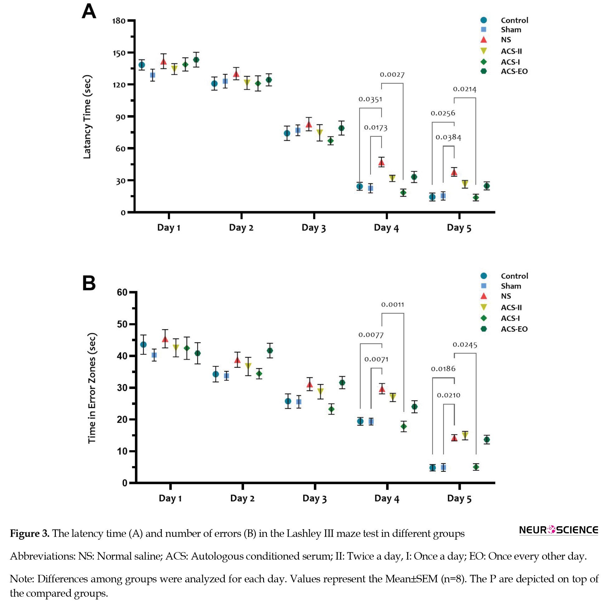
The latency time and number of errors were significantly increased in the NS group on days 4 and 5 (P<0.05) compared to the control and sham groups. In the ACS-I group, both indices were significantly reduced on the same days (at least P<0.05) (Figures 3A and 3B).
ACS effect on spatial memory in the Lashley III maze test
The latency time and number of errors were significantly increased in the NS group on days 4 and 5 (P<0.05) compared to the control and sham groups. In the ACS-I group, both indices were significantly reduced on the same days (at least P<0.05) (Figures 3A and 3B).
ACS increases IGF-1 and IL-1RA levels after mPFC ischemia
Quantitative analysis revealed significant reductions in both IGF-1 and IL-1RA levels in the NS-treated ischemic group compared to the control and sham groups (P<0.05). The ACS-I and ACS-EO groups exhibited an increase in IGF-1 expression; however, an increase in IL-1RA levels was observed only in the ACS-I group (at least P<0.01) (Figures 4A and 4C).
No significant differences were observed between the Sham and control groups for either biomarker.
ACS attenuates neuroinflammation and tau pathology following mPFC ischemia
Focal ischemia in the mPFC significantly elevated IL-1β levels and increased the p-tau to t-tau ratio compared to both control and sham groups (P<0.05). Treatment with ACS in all three groups significantly reduced both parameters (at least P<0.05) (Figurers 4B and 4D).
ACS reduces the mPFC infarct volume
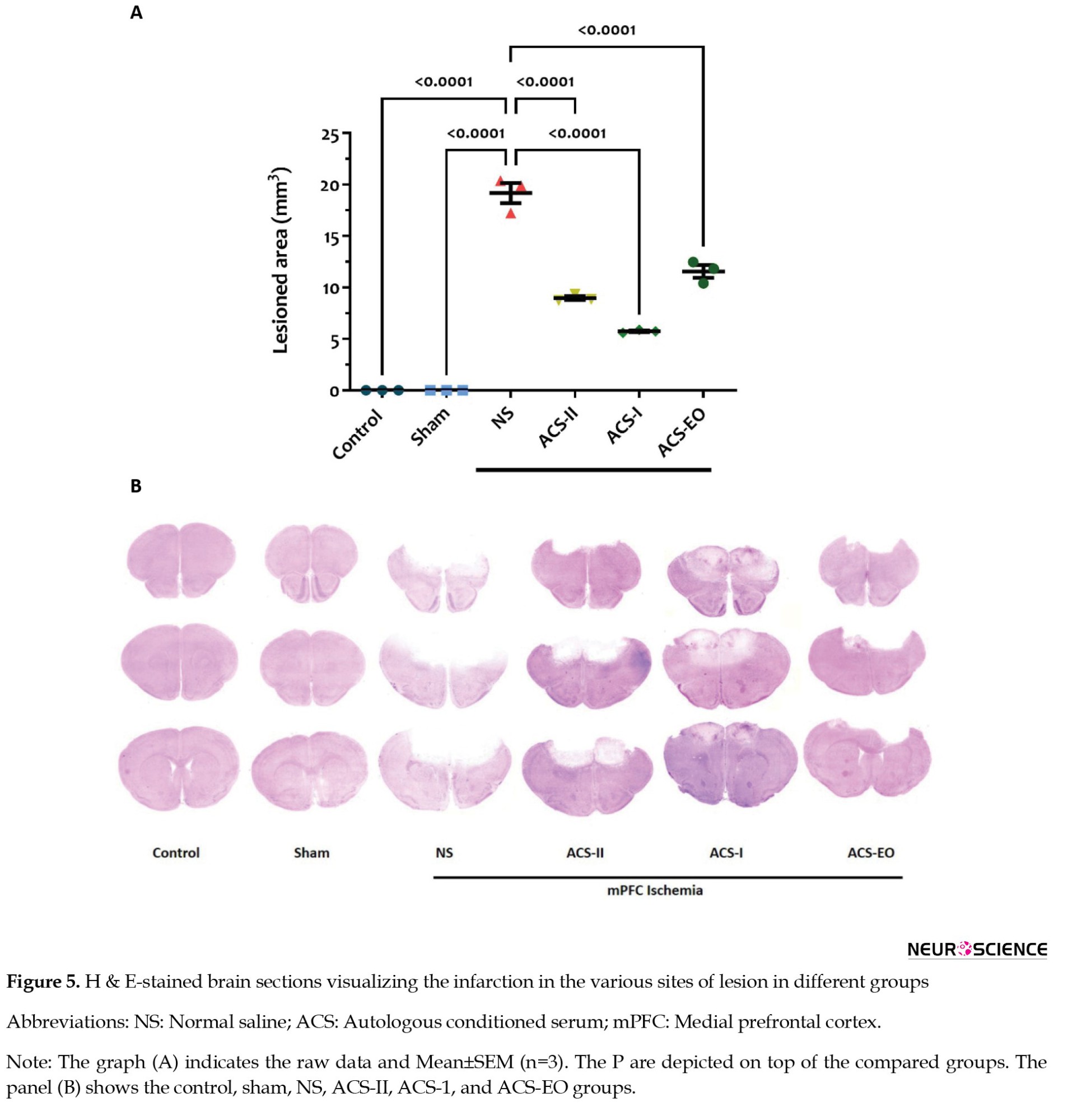
Ischemia induced infarction in the mPFC region, while ACS treatment in all three groups significantly reduced the infarct volume (P<0.0001) (Figure 5).
ACS attenuates neuroinflammation and tau pathology following mPFC ischemia
Focal ischemia in the mPFC significantly elevated IL-1β levels and increased the p-tau to t-tau ratio compared to both control and sham groups (P<0.05). Treatment with ACS in all three groups significantly reduced both parameters (at least P<0.05) (Figurers 4B and 4D).
ACS reduces the mPFC infarct volume
Ischemia induced infarction in the mPFC region, while ACS treatment in all three groups significantly reduced the infarct volume (P<0.0001) (Figure 5).
4. Discussion
According to the results of the present study, intranasal administration of ACS improves spatial and social memory impairments in mPFC ischemia in mice. The observed therapeutic effects likely result from dual mechanisms: Upregulation of neuroprotective IGF-1 and downregulation of pathological tau phosphorylation (p-tau/t-tau ratio) in peri-infarct areas. Additionally, ACS reduced pro-inflammatory cytokine (IL-1β) and augmented the level of IL-1RA cytokine in the ischemic area.
The global burden of stroke and dementia is rising at an alarming rate, with current epidemiological projections estimating that by 2050, there will be approximately 200 million stroke survivors and 106 million individuals living with dementia worldwide. This dramatic increase, representing a 150% surge in stroke prevalence and near-tripling of dementia cases compared to 2020 baselines, reflects the compounding effects of aging populations, particularly in low- and middle-income countries, alongside insufficient management of vascular risk factors and limited therapeutic breakthroughs for neurodegenerative processes (Brainin et al., 2020). Accordingly, it has been reported that approximately one in every three or four stroke survivors develops some form of cognitive decline or dementia (Kalaria et al., 2016), which hinders successful recovery. Clinical and imaging data have identified that functional outcomes following a stroke depend on factors such as the type of stroke, its severity, location, number of occurrences, and the volume of insult (Onyike, 2006; Rost et al., 2022). In this context, no significant differences have been reported in cognitive assessment between stroke subtypes (Aam et al., 2020); however, greater survival and incidence rates of ischemic stroke compared to hemorrhagic stroke leave a higher number of ischemic stroke survivors experiencing cognitive decline (Perna & Temple, 2015). Additionally, the anatomical location of the lesion is considered an influential factor for cognitive decline and is a strategic contributor to developing future dementia in stroke patients (Munsch et al., 2016; Zhao et al., 2017).
The PFC primarily contributes to a wide range of cognitive and executive functions, which are governed by lateral, medial, and orbitofrontal subregions exerting top-down control over other cortical and subcortical domains (Jobson et al., 2021). Data from electrophysiological and behavioral analyses suggest that the mPFC is implicated in high cognitive functions, including spatial working memory, attention, social behavior, and decision making (Carboni et al., 2024; Giacometti Giordani et al., 2023; Wirt & Hyman, 2017). Studies utilizing experimental models have indicated impaired recognition memory, spatial memory, cognitive inflexibility, and anxiety-related behavior following mPFC ischemia (Happ et al., 2020; Livingston-Thomas et al., 2015). Results from the current investigation revealed that mPFC ischemia increased latency and time spent in the error zone in the Lashley III maze, indicating impaired spatial learning. Similarly, it has been documented that focal ischemia in mPFC decreased displacement index in the what-where-which test, demonstrating impaired episodic memory (Sadigh-Eteghad et al., 2018). Moreover, the mPFC inactivation exhibited impaired inhibition of retrieval of non-practiced items, which is an essential phenomenon for successful memory recall by blocking retrieval of inappropriate competing interfaces (Wu et al., 2014). Scott et al. demonstrated that the inactivation of the mPFC led to a decline in odor working memory, and disconnecting its circuit with the mediodorsal thalamus resulted in dysfunction in exploratory motor activity (Scott et al., 2020).
We also found that mPFC photothrombotic ischemic induction dramatically declines the social memory index in the social interaction test. The mPFC has been proposed as a crucial foundation for social memory and social novelty encoding, facilitated by its extensive projections to limbic structures and top-down control over various circuits (Ko, 2017). The pharmacological blockade of the mPFC has been shown to block social recognition memory (Marcondes et al., 2020). Moreover, the long-term excitatory-inhibitory imbalance of hippocampal projections to mPFC has been found to impair social memory without affecting other social interactions, suggesting that any disruptions in the neural substrates of this circuit interfere with social memory processing (Phillips et al., 2019). It has been demonstrated that mPFC parvalbumin neurons mediate social behavior by targeting the hippocampal-dependent social memory pathway (Sun et al., 2020).
The IGF-1 is a trophic signal promoting maturation and growth in most tissues. Data from clinical studies have proven the positive role of IGF-1 overexpression in human motor and cognitive performances, while its suppression is observed in individuals with AD (Ferreira, 2021). Downregulation of IGF-1 compromised its capacity to promote the polarization of M2 microglia, suppressed microglia reactive oxygen species, and M1 phenotype markers like TNF-α, and inhibited astrocytic response to stimuli (Labandeira-Garcia et al., 2017). Furthermore, systemic inflammation has been shown to increase levels of the inflammatory cytokine TNF-α and IGF binding protein-1 (IGFBP-1), while reducing IGF-1 levels by approximately half in affected tissues, such as the brain (Fan et al., 1995). Additionally, intravenous IGF-1 improves the cognitive and neurological functions of rats subjected to ischemic stroke. This improvement was associated with the restoration of IGF-1 levels in the hippocampus, cortex, and amygdala of the affected rats. This exogenously increased levels of IGF-1 led to decreased levels of IL-6, IL-1β, and TNF-α in the plasma and hippocampus, as well as down-regulation of phospho-tau protein in the hippocampus and reduced cortical infarct volume (Yang et al., 2020).
P-tau is considered a shared biomarker between stroke and neurodegenerative disease. The higher plasma level of p-tau in patients with a history of stroke, regardless of whether they have dementia or not, compared to control individuals (Tang et al., 2018), could be related to the blood-brain barrier disruption following a stroke (Kurzepa et al., 2010). Subcortical focal ischemia was proposed to upregulate the microtubule-associated regulatory kinase, leading to microtubule destabilization by phosphorylation of tau at serine-262 in the microtubule-binding domain (Hayden et al., 2019). Tau phosphorylation at this site is believed to act as a gateway phosphorylation site to enhance further phosphorylation and tau aggregation (Biernat et al., 1993). Neuroinflammation as a pathological hallmark of ischemic stroke develops within minutes and continues for days and weeks following the attack (Anrather & Iadecola, 2016). Cells in the ischemic core and peri-infarct zone begin to release inflammatory molecules such as cytokines and chemokines into the systemic circulation, leading to the infiltration of peripheral immune cells into the brain, which then activate the host immune system (Wu et al., 2022; Xu et al., 2020). The microglia express pro- and anti-inflammatory cytokines in infarct and peri-infarct lesion sites (Lambertsen et al., 2019; Lambertsen et al., 2019).
IL-1RA endogenously inhibits IL-1α and IL-1β, working by blocking the IL-1β signaling pathway to reduce inflammation (Harrell et al., 2020). Due to the low concentration of IL-1RA in affected tissues, boosting the level of IL-1RA could be an appealing target to achieve maximum therapeutic benefits (Camargo Garbin & Morris, 2021). Recently, immunotherapy in clinical stroke trials has gained attention for targeting innate and adaptive responses by lowering microglia activation, suppressing immune cell migration to the brain, and modulating the IL-1β signaling pathway (Drieu et al., 2018). ACS is a bioproduct enriched in IL-1RA, IL-4, IL-10, and growth factors (Shakouri et al., 2021), which has been clinically used in orthopedic disorders and is currently in clinical trials for COVID-19 (Fotouhi et al., 2018; Shakouri et al., 2020). In the present investigation, we demonstrated that intranasal ACS therapy buffered spatial memory and social memory dysfunctions, which probably occurred through its anti-inflammatory abilities to upregulate IGF-1 and IL-1RA and downregulate IL-1β and p-tau in the mPFC of animals that underwent photothrombotic ischemic stroke. Inhibiting IL-1RA has been claimed to increase vascular inflammation by increasing IL-1 signaling, introducing it as an anti-inflammatory therapeutic option in conditions such as ischemic stroke. In this context, it has been reported that blocking IL-1α after stroke reduced endothelial activation and expression of adhesion molecules. This condition led to a decrease in penumbral mononuclear phagocyte content and neurotoxic mediators like matrix metalloprotease, ultimately modulating cerebral injury (Liberale et al., 2021). In agreement, data from a phase II placebo-controlled clinical trial in ischemic and hemorrhagic stroke cases has shown that the subcutaneous injection of recombinant human IL-1RA reduced the plasma level of IL-6 and C-reactive protein and also decreased the extent of edema observed in CT scans of hemorrhagic stroke patients (Parry-Jones et al., 2023; Smith et al., 2018). It is suggested that IL-1RA enhanced the effectiveness of tissue plasminogen activator (tPA) treatment, a gold standard therapy for ischemic stroke, by inhibiting IL-1 to elevate the levels of endogenous tPA inhibitor-1 (Smith et al., 2018). It has been accepted that microglia IL-1β increases neuronal tau phosphorylation through p38-MAPk signaling pathway, thereby disrupt cytoskeleton assembly and axon stabilization leading to neuronal cell death (Li et al., 2003). Indeed, IL-1β serves as a vital substrate for tauopathy conditions’ pathological biomarkers (Maphis et al., 2015). IL-1β-treated microglial culture has been shown to upregulate the level of secreted fragment of the β-amyloid precursor protein (s-APP). Given the positive role of pre-treatment with conditioned medium from sAPP-activated microglia with IL-RA in down-regulation of of β- p-tau, α-synuclein, and p38-MAPk, IL-1R could be assigned as a promising therapy in tauopathy conditions (Griffin et al., 2006).
5. Conclusion
The data from our study showed that chronic ACS administration after stroke significantly improved spatial and social interaction memories. Additionally, ACS therapy notably increased the protein abundance of IGF-1 and IL-1RA, while simultaneously decreasing the production of IL-1β and p-tau in the mPFC. However, to fully elucidate the cellular and molecular mechanisms, further exact studies are needed to examine how ACS mediates the observed neuroprotective effects against brain ischemia.
Ethical Considerations
Compliance with ethical guidelines
This study was approved by the Ethics Committee of Tabriz University of Medical Sciences, Tabriz, Iran (Code: IR.TBZMED.VCR.REC.1399.403) and performed following the guidelines of the National Institutes of Health (NIH) (Publication No. 85–23, revised 1985).
Funding
This research was supported by a grant from the Tabriz University of Medical sciences, Tabriz, Iran (Grant No.: 60948).
Authors' contributions
Conceptualization: Sareh Kazmi, Alireza Pishgahi, Mehdi Yousefi, Tahereh Ghadiri, Saeed Sadigh-Eteghad, and Saeed Sadigh-Eteghad; Investigation: Sareh Kazmi, Fatemehsadat Seyedaghamiri, Alireza Pishgahi and Mehdi Yousefi: Data curation: Sareh Kazmi; Formal analysis: Sareh Kazmi and Saeed Sadigh-Eteghad; Validation: Saeed Sadigh-Eteghad; Writing: Sareh Kazmi, Neda Yazdanfar, Tahereh Ghadiri, Saeed Sadigh-Eteghad, and Mehdi Farhoudi; final approval: All authors.
Conflict of interest
The authors declared no conflict of interest.
Acknowledgments
The authors would like to appreciate the Tabriz University of Medical sciences for supporting of this work.
References
Aam, S., Einstad, M. S., Munthe-Kaas, R., Lydersen, S., Ihle-Hansen, H., & Knapskog, A. B., et al. (2020). Post-stroke cognitive impairment-impact of follow-up time and stroke subtype on severity and cognitive profile: The Nor-COAST study. Frontiers in Neurology, 11, 699. [DOI:10.3389/fneur.2020.00699] [PMID]
Angadi, D. S., Macdonald, H., & Atwal, N. (2020). Autologous cell-free serum preparations in the management of knee osteoarthritis: What is the current clinical evidence? Knee Surgery & Related Research, 32(1), 16. [DOI:10.1186/s43019-020-00036-5] [PMID]
Anrather, J., & Iadecola, C. (2016). Inflammation and stroke: An overview. Neurotherapeutics, 13(4), 661-670. [DOI:10.1007/s13311-016-0483-x] [PMID]
Bhasin, A., Srivastava, M., Bhatia, R., Mohanty, S., Kumaran, S., & Bose, S. (2012). Autologous intravenous mononuclear stem cell therapy in chronic ischemic stroke. Journal of Stem Cells & Regenerative Medicine, 8(3), 181-189. [DOI:10.46582/jsrm.0803011] [PMID]
Bi, M., Gladbach, A., van Eersel, J., Ittner, A., Przybyla, M., & van Hummel, A., et al. (2017). Tau exacerbates excitotoxic brain damage in an animal model of stroke. Nature Communications, 8(1), 473. [DOI:10.1038/s41467-017-00618-0] [PMID]
Biernat, J., Gustke, N., Drewes, G., Mandelkow, E. M., & Mandelkow, E. (1993). Phosphorylation of Ser262 strongly reduces binding of tau to microtubules: distinction between PHF-like immunoreactivity and microtubule binding. Neuron, 11(1), 153–163. [DOI:10.1016/0896-6273(93)90279-z] [PMID]
Brainin, M., Feigin, V. L., Norrving, B., Martins, S. C. O., Hankey, G. J., & Hachinski, V., et al. (2020). Global prevention of stroke and dementia: The WSO Declaration. The Lancet Neurology, 19(6), 487-488. [DOI:10.1016/S1474-4422(20)30141-1] [PMID]
Brassai, A., Suvanjeiev, R. G., Bán, E. G., & Lakatos, M. (2015). Role of synaptic and nonsynaptic glutamate receptors in ischaemia induced neurotoxicity. Brain Research Bulletin, 112, 1-6. [DOI:10.1016/j.brainresbull.2014.12.007] [PMID]
Camargo Garbin, L., & Morris, M. J. (2021). A comparative review of autologous conditioned serum and autologous protein solution for treatment of osteoarthritis in horses. Frontiers in Veterinary Science, 8, 602978. [DOI:10.3389/fvets.2021.602978] [PMID]
Carboni, E., Ibba, M., Carboni, E., & Carta, A. R. (2024). Adolescent stress differentially modifies dopamine and norepinephrine release in the medial prefrontal cortex of adult rats. Progress in Neuro-Psychopharmacology and Biological Psychiatry, 134, 111055. [DOI:10.1016/j.pnpbp.2024.111055] [PMID]
Chen, J., Li, Y., Wang, L., Zhang, Z., Lu, D., & Lu, M., et al. (2001). Therapeutic benefit of intravenous administration of bone marrow stromal cells after cerebral ischemia in rats. Stroke, 32(4), 1005-1011. [DOI:10.1161/01.str.32.4.1005] [PMID]
Chen, J., Sanberg, P. R., Li, Y., Wang, L., Lu, M., & Willing, A. E., et al. (2001). Intravenous administration of human umbilical cord blood reduces behavioral deficits after stroke in rats. Stroke, 32(11), 2682–2688. [DOI:10.1161/hs1101.098367] [PMID]
Chen, X., & Jiang, H. (2019). Tau as a potential therapeutic target for ischemic stroke. Aging, 11(24), 12827–12843. [DOI:10.18632/aging.102547] [PMID]
Doeppner, T. R., Herz, J., Görgens, A., Schlechter, J., Ludwig, A. K., & Radtke, S., et al. (2015). Extracellular vesicles improve post-stroke neuroregeneration and prevent postischemic immunosuppression. Stem Cells Translational Medicine, 4(10), 1131–1143. [DOI:10.5966/sctm.2015-0078] [PMID]
Drieu, A., Levard, D., Vivien, D., & Rubio, M. (2018). Anti-inflammatory treatments for stroke: From bench to bedside. Therapeutic Advances in Neurological Disorders, 11, 1756286418789854. [DOI:10.1177/1756286418789854] [PMID]
Fan, J., Li, Y. H., Bagby, G. J., & Lang, C. H. (1995). Modulation of inflammation-induced changes in insulin-like growth factor (IGF)-I and IGF binding protein-1 by anti-TNF antibody. Shock, 4(1), 21-26. [DOI:10.1097/00024382-199507000-00003] [PMID]
Ferreira, S. T. (2021). Brain insulin, insulin‐like growth factor 1 and glucagon‐like peptide 1 signalling in Alzheimer’s disease. Journal of Neuroendocrinology, 33(4), e12959. [DOI:10.1111/jne.12959] [PMID]
Fotouhi, A., Maleki, A., Dolati, S., Aghebati-Maleki, A., & Aghebati-Maleki, L. (2018). Platelet rich plasma, stromal vascular fraction and autologous conditioned serum in treatment of knee osteoarthritis. Biomedicine & Pharmacotherapy, 104, 652–660. [DOI:10.1016/j.biopha.2018.05.019] [PMID]
Frizziero, A., Giannotti, E., Oliva, F., Masiero, S., & Maffulli, N. (2013). Autologous conditioned serum for the treatment of osteoarthritis and other possible applications in musculoskeletal disorders. British Medical Bulletin, 105, 169–184. [DOI:10.1093/bmb/lds016] [PMID]
Giacometti Giordani, L., Crisafulli, A., Cantarella, G., Avenanti, A., & Ciaramelli, E. (2023). The role of posterior parietal cortex and medial prefrontal cortex in distraction and mind-wandering. Neuropsychologia, 188, 108639. [PMID]
Gong, P., Zou, Y., Zhang, W., Tian, Q., Han, S., & Xu, Z., et al. (2021). The neuroprotective effects of insulin-like growth factor 1 via the Hippo/YAP signaling pathway are mediated by the PI3K/AKT cascade following cerebral ischemia/reperfusion injury. Brain Research Bulletin, 177, 373-387. [DOI:10.1016/j.brainresbull.2021.10.017] [PMID]
Griffin, W. S., Liu, L., Li, Y., Mrak, R. E., & Barger, S. W. (2006). Interleukin-1 mediates Alzheimer and Lewy body pathologies. Journal of Neuroinflammation, 3, 5. [DOI:10.1186/1742-2094-3-5] [PMID]
Happ, D. F., Wegener, G., & Tasker, R. A. (2020). Effect of ischemic lesions in medial prefrontal cortex and nucleus accumbens on affective behavior in rats. Behavioural Brain Research, 378, 112234. [DOI:10.1016/j.bbr.2019.112234] [PMID]
Harrell, C. R., Markovic, B. S., Fellabaum, C., Arsenijevic, N., Djonov, V., & Volarevic, V. (2020). The role of Interleukin 1 receptor antagonist in mesenchymal stem cell-based tissue repair and regeneration. BioFactors (Oxford, England), 46(2), 263–275. [DOI:10.1002/biof.1587] [PMID]
Hayden, E. Y., Putman, J., Nunez, S., Shin, W. S., Oberoi, M., & Charreton, M., et al. (2019). Ischemic axonal injury up-regulates MARK4 in cortical neurons and primes tau phosphorylation and aggregation. Acta Neuropathologica Communications, 7(1), 135. [DOI:10.1186/s40478-019-0783-6] [PMID]
Houlton, J., Barwick, D., & Clarkson, A. N. (2021). Frontal cortex stroke-induced impairment in spatial working memory on the trial-unique nonmatching-to-location task in mice. Neurobiology of Learning and Memory, 177, 107355. [DOI:10.1016/j.nlm.2020.107355] [PMID]
Jobson, D. D., Hase, Y., Clarkson, A. N., & Kalaria, R. N. (2021). The role of the medial prefrontal cortex in cognition, ageing and dementia. Brain Communications, 3(3), fcab125.[DOI:10.1093/braincomms/fcab125] [PMID]
Kalaria, R. N., Akinyemi, R., & Ihara, M. (2016). Stroke injury, cognitive impairment and vascular dementia. Biochimica et Biophysica Acta, 1862(5), 915–925. [DOI:10.1016/j.bbadis.2016.01.015] [PMID]
Ko, J. (2017). Neuroanatomical substrates of rodent social behavior: The medial prefrontal cortex and its projection patterns. Frontiers in Neural Circuits, 11, 41. [DOI:10.3389/fncir.2017.00041] [PMID]
Kurzepa, J., Bielewicz, J., Grabarska, A., Stelmasiak, Z., Stryjecka-Zimmer, M., & Bartosik-Psujek, H. (2010). Matrix metalloproteinase-9 contributes to the increase of tau protein in serum during acute ischemic stroke. Journal of Clinical Neuroscience, 17(8), 997-999. [DOI:10.1016/j.jocn.2010.01.005] [PMID]
Labandeira-Garcia, J. L., Costa-Besada, M. A., Labandeira, C. M., Villar-Cheda, B., & Rodríguez-Perez, A. I. (2017). Insulin-like growth factor-1 and neuroinflammation. Frontiers in Aging Neuroscience, 9, 365. [DOI:10.3389/fnagi.2017.00365] [PMID]
Lambertsen, K. L., Finsen, B., & Clausen, B. H. (2019). Post-stroke inflammation-target or tool for therapy? Acta Neuropathologica, 137(5), 693-714. [DOI:10.1007/s00401-018-1930-z] [PMID]
Li, W., Ye, A., Ao, L., Zhou, L., Yan, Y., & Hu, Y., et al. (2020). Protective mechanism and treatment of neurogenesis in cerebral ischemia. Neurochemical Research, 45(10), 2258-2277. [DOI:10.1007/s11064-020-03092-1] [PMID]
Li, Y., Liu, L., Barger, S. W., & Griffin, W. S. (2003). Interleukin-1 mediates pathological effects of microglia on tau phosphorylation and on synaptophysin synthesis in cortical neurons through a p38-MAPK pathway. The Journal of Neuroscience: The Official Journal of the Society for Neuroscience, 23(5), 1605–1611. [DOI:10.1523/jneurosci.23-05-01605.2003] [PMID]
Liberale, L., Ministrini, S., Carbone, F., Camici, G. G., & Montecucco, F. (2021). Cytokines as therapeutic targets for cardio- and cerebrovascular diseases. Basic Research in Cardiology, 116(1), 23. [DOI:10.1007/s00395-021-00863-x] [PMID]
Livingston-Thomas, J. M., Jeffers, M. S., Nguemeni, C., Shoichet, M. S., Morshead, C. M., & Corbett, D. (2015). Assessing cognitive function following medial prefrontal stroke in the rat. Behavioural Brain Research, 294, 102–110. [DOI:10.1016/j.bbr.2015.07.053] [PMID]
Maphis, N., Xu, G., Kokiko-Cochran, O. N., Jiang, S., Cardona, A., & Ransohoff, R. M., et al. (2015). Reactive microglia drive tau pathology and contribute to the spreading of pathological tau in the brain. Brain, 138(Pt 6), 1738-1755. [DOI:10.1093/brain/awv081] [PMID]
Marcondes, L. A., Nachtigall, E. G., Zanluchi, A., de Carvalho Myskiw, J., Izquierdo, I., & Furini, C. R. G. (2020). Involvement of medial prefrontal cortex NMDA and AMPA/kainate glutamate receptors in social recognition memory consolidation. Neurobiology of Learning and Memory, 168, 107153. [DOI:10.1016/j.nlm.2019.107153] [PMID]
Munsch, F., Sagnier, S., Asselineau, J., Bigourdan, A., Guttmann, C. R., & Debruxelles, S., et al. (2016). Stroke location is an independent predictor of cognitive outcome. Stroke, 47(1), 66-73. [DOI:10.1161/STROKEAHA.115.011242] [PMID]
Onyike, C. U. (2006). Cerebrovascular disease and dementia. International Review of Psychiatry (Abingdon, England), 18(5), 423–431. [DOI:10.1080/09540260600935421] [PMID]
Parry-Jones, A. R., Stocking, K., MacLeod, M. J., Clarke, B., Werring, D. J., & Muir, K. W., et al. (2023). Phase II randomised, placebo-controlled, clinical trial of interleukin-1 receptor antagonist in intracerebral haemorrhage: BLOcking the cytokine IL-1 in ICH (BLOC-ICH). European Stroke Journal, 8(3), 819–827.[DOI:10.1177/23969873231185208] [PMID]
Perna, R., & Temple, J. (2015). Rehabilitation outcomes: Ischemic versus hemorrhagic strokes. Behavioural Neurology, 2015, 891651. [DOI:10.1155/2015/891651] [PMID]
Phillips, M. L., Robinson, H. A., & Pozzo-Miller, L. (2019). Ventral hippocampal projections to the medial prefrontal cortex regulate social memory. Elife, 8, e44182. [DOI:10.7554/eLife.44182] [PMID]
Pluta, R., Czuczwar, S. J., Januszewski, S., & Jabłoński, M. (2021). The many faces of post-ischemic tau protein in brain neurodegeneration of the Alzheimer’s disease type. Cells, 10(9), 2213. [DOI:10.3390/cells10092213] [PMID]
Pluta, R., Kiś, J., Januszewski, S., Jabłoński, M., & Czuczwar, S. J. (2022). Cross-talk between amyloid, tau protein and free radicals in post-ischemic brain neurodegeneration in the form of Alzheimer’s disease proteinopathy. Antioxidants, 11(1), 146. [DOI:10.3390/antiox11010146] [PMID]
Rost, N. S., Brodtmann, A., Pase, M. P., van Veluw, S. J., Biffi, A., & Duering, M., et al. (2022). Post-stroke cognitive impairment and dementia. Circulation Research, 130(8), 1252-1271. [DOI:10.1161/CIRCRESAHA.122.319951] [PMID]
Sadigh-Eteghad, S., Geranmayeh, M. H., Majdi, A., Salehpour, F., Mahmoudi, J., & Farhoudi, M. (2018). Intranasal cerebrolysin improves cognitive function and structural synaptic plasticity in photothrombotic mouse model of medial prefrontal cortex ischemia. Neuropeptides, 71, 61-69. [DOI:10.1016/j.npep.2018.07.002] [PMID]
Scott, G. A., Liu, M. C., Tahir, N. B., Zabder, N. K., Song, Y., & Greba, Q., et al. (2020). Roles of the medial prefrontal cortex, mediodorsal thalamus, and their combined circuit for performance of the odor span task in rats: Analysis of memory capacity and foraging behavior. Learning & Memory, 27(2), 67-77. [DOI:10.1101/lm.050195.119] [PMID]
Seyedaghamiri, F., Farajdokht, F., Vatandoust, S. M., Mahmoudi, J., Khabbaz, A., & Sadigh-Eteghad, S. (2021). Sericin modulates learning and memory behaviors by tuning of antioxidant, inflammatory, and apoptotic markers in the hippocampus of aged mice. Molecular Biology Reports, 48(2), 1371-1382. [DOI:10.1007/s11033-021-06195-2] [PMID]
Shakouri, S. K., Dolati, S., Santhakumar, J., Thakor, A. S., & Yarani, R. (2021). Autologous conditioned serum for degenerative diseases and prospects. Growth Factors, 39(1-6), 59-70. [DOI:10.1080/08977194.2021.2012467] [PMID]
Shakouri, S. K., Roshangar, L., & Mahmoodpoor, A. (2020). Intratracheal administration of autologus conditioned serum for COVID-19 associated respiratory distress syndrome. Journal of Critical Care, 60, 209–211. [DOI:10.1016/j.jcrc.2020.08.016] [PMID]
Smith, C. J., Hulme, S., Vail, A., Heal, C., Parry-Jones, A. R., & Scarth, S., et al. (2018). SCIL-STROKE (subcutaneous interleukin-1 receptor antagonist in ischemic stroke): A randomized controlled phase 2 trial. Stroke, 49(5), 1210-1216. [DOI:10.1161/STROKEAHA.118.020750] [PMID]
Sun, Q., Li, X., Li, A., Zhang, J., Ding, Z., & Gong, H., et al. (2020). Ventral hippocampal-prefrontal interaction affects social behavior via parvalbumin positive neurons in the medial prefrontal cortex. iScience, 23(3), 100894. [DOI:10.1016/j.isci.2020.100894] [PMID]
Tang, S. C., Yang, K. C., Chen, C. H., Yang, S. Y., Chiu, M. J., & Wu, C. C., et al. (2018). Plasma β-amyloids and tau proteins in patients with vascular cognitive impairment. Neuromolecular Medicine, 20(4), 498–503. [DOI:10.1007/s12017-018-8513-y] [PMID]
Uchihara, T., Nakamura, A., Arai, T., Ikeda, K., & Tsuchiya, K. (2004). Microglial tau undergoes phosphorylation-independent modification after ischemia. Glia, 45(2), 180-187. [DOI:10.1002/glia.10318] [PMID]
Wehling, P., Moser, C., Frisbie, D., Wayne McIlwraith, C., Kawcak, C. E., & Krauspe, R., et al. (2007). Autologous conditioned serum in the treatment of orthopedic diseases. BioDrugs Clinical Immunotherapeutics, Biopharmaceuticals and Gene Therapy, 21(5), 323–332. [DOI:10.2165/00063030-200721050-00004] [PMID]
Wirt, R. A., & Hyman, J. M. (2017). Integrating spatial working memory and remote memory: Interactions between the medial prefrontal cortex and hippocampus. Brain Sciences, 7(4), 43. [DOI:10.3390/brainsci7040043] [PMID]
Wu, F., Liu, Z., Zhou, L., Ye, D., Zhu, Y., & Huang, K., et al. (2022). Systemic immune responses after ischemic stroke: From the center to the periphery. Frontiers in Immunology, 13, 911661.[DOI:10.3389/fimmu.2022.911661] [PMID]
Wu, J. Q., Peters, G. J., Rittner, P., Cleland, T. A., & Smith, D. M. (2014). The hippocampus, medial prefrontal cortex, and selective memory retrieval: Evidence from a rodent model of the retrieval‐induced forgetting effect. Hippocampus, 24(9), 1070-1080. [DOI:10.1002/hipo.22291] [PMID]
Xu, S., Lu, J., Shao, A., Zhang, J. H., & Zhang, J. (2020). Glial cells: Role of the immune response in ischemic stroke. Frontiers in Immunology, 11, 294. [DOI:10.3389/fimmu.2020.00294] [PMID]
Yang, J., Wu, S., Hou, L., Zhu, D., Yin, S., & Yang, G., et al. (2020). Therapeutic effects of simultaneous delivery of nerve growth factor mRNA and protein via exosomes on cerebral ischemia. Molecular Therapy. Nucleic Acids, 21, 512–522. [DOI:10.1016/j.omtn.2020.06.013] [PMID]
Yang, W., Li, G., Cao, K., Ma, P., Guo, Y., & Tong, W., et al. (2020). Exogenous insulin-like growth factor 1 attenuates acute ischemic stroke-induced spatial memory impairment via modulating inflammatory response and tau phosphorylation. Neuropeptides, 83, 102082. [DOI:10.1016/j.npep.2020.102082] [PMID]
Zhao, L., Biesbroek, J. M., Shi, L., Liu, W., Kuijf, H. J., & Chu, W. W., et al. (2018). Strategic infarct location for post-stroke cognitive impairment: A multivariate lesion-symptom mapping study. Journal of Cerebral Blood Flow & Metabolism, 38(8), 1299-1311. [DOI:10.1177/0271678X17728162] [PMID]
Zhou, J., Du, T., Li, B., Rong, Y., Verkhratsky, A., & Peng, L. (2015). Crosstalk between MAPK/ERK and PI3K/AKT signal pathways during brain ischemia/reperfusion. ASN Neuro, 7(5), 1759091415602463. [DOI:10.1177/1759091415602463] [PMID]
Type of Study: Original |
Subject:
Cellular and molecular Neuroscience
Received: 2025/03/3 | Accepted: 2025/05/12 | Published: 2025/07/1
Received: 2025/03/3 | Accepted: 2025/05/12 | Published: 2025/07/1
Send email to the article author
| Rights and permissions | |
 |
This work is licensed under a Creative Commons Attribution-NonCommercial 4.0 International License. |

Copyright © The Author(s);
This is an open access article distributed under the terms of the Creative Commons Attribution License (CC-By-NC), which permits use, distribution, and reproduction in any medium, provided the original work is properly cited and is not used for commercial purposes.
Contact Information