

Volume 17, Issue 2 (March & April 2026)
BCN 2026, 17(2): 323-336 |
Back to browse issues page


Download citation:
BibTeX | RIS | EndNote | Medlars | ProCite | Reference Manager | RefWorks
Send citation to:


BibTeX | RIS | EndNote | Medlars | ProCite | Reference Manager | RefWorks
Send citation to:
Golmohammadi H, Ghalandari-Shamami M, Haghparast A. Comparing the Effects of the Intra-DG Administration of Dopamine Receptor Antagonists’ Effects on Stress-induced Antinociception in Wistar Rats. BCN 2026; 17 (2) :323-336
URL: http://bcn.iums.ac.ir/article-1-3493-en.html
URL: http://bcn.iums.ac.ir/article-1-3493-en.html


1- Neuroscience Research Center, Institute of Neuroscience and Cognition, Shahid Beheshti University of Medical Sciences, Tehran, Iran.
2- Department of Physiology, TeMS.C., Islamic Azad University, Tehran, Iran. & Cognitive and Neuroscience Research Center (CNRC), TeMS.C., Islamic Azad University, Tehran, Iran.
3- Neuroscience Research Center, Institute of Neuroscience and Cognition, Shahid Beheshti University of Medical Sciences, Tehran, Iran. & School of Cognitive Sciences, Institute for Research in Fundamental Sciences, Tehran, Iran. & Department of Basic Sciences, Iranian Academy of Medical Sciences, Tehran, Iran. & National Brain Mapping Lab, Tehran, Iran.
2- Department of Physiology, TeMS.C., Islamic Azad University, Tehran, Iran. & Cognitive and Neuroscience Research Center (CNRC), TeMS.C., Islamic Azad University, Tehran, Iran.
3- Neuroscience Research Center, Institute of Neuroscience and Cognition, Shahid Beheshti University of Medical Sciences, Tehran, Iran. & School of Cognitive Sciences, Institute for Research in Fundamental Sciences, Tehran, Iran. & Department of Basic Sciences, Iranian Academy of Medical Sciences, Tehran, Iran. & National Brain Mapping Lab, Tehran, Iran.
Keywords: Restraint stress (RS), Force swimming stress, Dentate gyrus (DG), Dopamine receptors, Tail-flick test, Rat
Full-Text [PDF 1945 kb]
| Abstract (HTML)
Locomotor activity
The total distance traveled by the subject in an open field (60×60 cm2) over 60 min was tracked using a 3-CCD camera (Panasonic Corporation, Osaka, Japan), and the data were analyzed by Ethovision software (version 7; Noldus Information Technology, Wageningen, The Netherlands).
Statistical analysis
Statistical analysis of data was performed using GraphPad Prism software, version 6 (GraphPad Software, San Diego, USA). Results were reported as Mean±SEM, and normality of data distribution was assessed using the Kolmogorov–Smirnov test. Two-way repeated measures ANOVA with Bonferroni post hoc test was used to compare the percentage of maximum possible effect at different times between treatment groups. Also, one-way ANOVA followed by Tukey’s post hoc test was used to compare groups, when necessary.
To evaluate the effect of RS and FSS on acute pain behaviors, the area under the curve (AUC) values of different groups were calculated, and the unpaired t-test was used to compare the groups. In all analyses, a statistical significance level of P<0.05 was considered.
Results
Effects of RS and FSS on antinociceptive behaviors in the tail-flick test
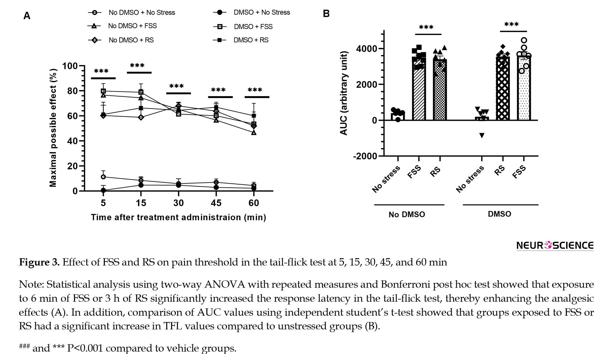
Pain-related behavior in the tail-flick test, as an animal model of acute pain, was assessed at time intervals of 5, 15, 30, 45, and 60 minutes, and the MPE% was obtained, and the AUC of the data related to TFL times after the application of the painful stimulus was examined. Two-way analysis of variance (ANOVA) followed by Bonferroni post-test showed that pain responses in the tail-flick test were significantly reduced in the groups exposed to RS and FSS compared to the non-stress groups (Figure 3A) [treatment effect: F5, 180=108.3, P<0.0001; time effect: F4, 180=3.731, P=0.006; treatment and time interaction effect: F20, 180=1.090, P=0.3629].
Discussion
The most important findings of the present investigation were as follows: (a) RS and FSS significantly increased antinociceptive behaviors in an acute model of pain compared to the control group; (b) intra-DG administration of SCH-23390 significantly reduced antinociceptive behaviors in the RS group compared to the FSS group; (c) the effect of FSS was more remarkable than that of RS on antinociceptive behaviors in groups that received sulpiride in the DG.
The DG, as part of the hippocampus, plays a key role in emotional processing, learning, and memory (Sun et al., 2023; Tyng et al., 2017). Kesner showed that the DG plays a fundamental role in relaying cortical inputs to the limbic system and in integrating emotional responses (Kesner, 2007). Since both pain and stress are associated with emotional processing, changes in dopamine receptor activity in the DG could directly influence the coordination of behavioral and neurochemical responses to stress.
The phenomenon of pain inhibition in response to stress is known as SIA (al’Absi et al., 2021). Exposure to stressful or fearful stimuli, whether conditioned or unconditioned, serves as a template for activating the endogenous pain inhibition system and can elicit this response (Taylor, 2017). The function of SIA is influenced by descending pain inhibition pathways, and any change in the activity of these pathways can disrupt it (Kwon et al., 2014). This suggests a complex and bidirectional interaction between stress and pain, in that pain can lead to stress, and stress can, depending on the circumstances, reduce or increase the intensity of pain (Melzack, 1999). The type of stressor determines the neural circuitry involved in pain inhibition. In general, mild stressors act primarily through mechanisms dependent on the endogenous opioid system, whereas severe stressors activate more opioid-independent pathways (Ferdousi & Finn, 2018). Furthermore, evidence suggests that different types of stressors can exert multifaceted and complex effects on dopamine levels in the mesolimbic system (Baik, 2020).
Several studies have shown that various forms of stress, including forced swimming, restraint, sleep deprivation, and food restriction or deprivation, can alter the threshold and intensity of pain perception (Butler & Finn, 2009). In the present study, RS and FSS were used as aversive stressors to assess the effect of stress on pain responses in rats. The findings showed that RS and FSS administration resulted in an analgesic effect in acute pain and significantly increased the latency to respond during a 60-min tail-flick test. The differential effect of sulpiride in various stress paradigms suggests that different neurochemical mechanisms are responsible for SIA, and this depends on the type of stressor. While D1 receptor signaling is dominant in RS, D2 pathways may be more activated in FSS, suggesting a specific involvement of dopaminergic circuits depending on the type of stressor. These results are consistent with previous reports that have confirmed the inhibitory effect of stress in animal models of thermal and visceral pain (Askari et al., 2021; Faramarzi et al., 2016).
SIA plays an important role in the temporary inhibition of nociceptive transmission as an adaptive mechanism in the face of environmental stressors and has attracted the attention of researchers to identify the central neural networks involved in pain inhibition (Butler & Finn, 2009). Although this phenomenon was initially thought to be mediated mainly by the endogenous opioid system, subsequent evidence has shown that non-opioid mechanisms also contribute significantly (Mogil et al., 1996). For example, in the dorsal hippocampus of the guinea pig, pain transmission has been shown to be regulated by the interaction of GABAergic, cholinergic, and opioidergic systems (Mendes & Menescal-de-Oliveira, 2008). Furthermore, during SIA, activation of a network of neurotransmitters and neuromodulators, including monoamines, GABA, glutamate, and endocannabinoids has been reported, indicating the multisystem and complex nature of this phenomenon (Butler & Finn, 2009).
Our results indicated that intra-DG administration of SCH-23390 significantly reduced antinociceptive behaviors in RS more than in FSS, but the effect of FSS was more remarkable than RS on antinociceptive behaviors in groups that received sulpiride in the DG.
The findings suggest that individual susceptibility to SIA is highly variable and is associated with a variety of characteristics, including the type and intensity of stress and the degree of opioid sensitivity and response to shocking stimuli. In addition, SIA is influenced by environmental and individual factors, such as age, gender, and previous painful or stressful experiences (Butler & Finn, 2009). In a recent study, the application of three hours of RS induced SIA in both the acute and inflammatory phases of the formalin test. However, when dopamine receptor antagonists were microinjected into the DG, the intensity of this analgesic response was significantly reduced in both phases of the test (Nazari-Serenjeh et al., 2024). These findings suggest that various kinds of stressors act in pain modulation in descending pathways through specific receptors. However, to more precisely elucidate the contribution of each of these receptor subtypes in the process of pain modulation, a comprehensive and integrated approach is necessary.
Evidence suggests that RS and FSS can induce different patterns of dopamine release in the DG and extended hippocampal circuits. RS, as a long-term psychological stimulus, often induces a sustained and moderate release of dopamine that activates mostly high-affinity D1 receptors. In contrast, FSS, which is short-term but highly physically demanding, can induce a rapid and phasic release of dopamine and more effectively activates D2 receptors, pathways that are associated with motivational and coping responses (Barrot et al., 2000, Kalivas & Duffy, 1995). RS primarily activates cortico-limbic afferents, including inputs from the PFC and amygdala (McKlveen et al., 2015), regions that are strongly modulated by D1 receptor-mediated dopaminergic signaling (Seamans & Yang, 2004). In contrast, FSS engages broader motivational and mesolimbic circuits, particularly the ventral tegmental area–nucleus accumbens pathway (Kalivas & Duffy, 1995; Russo & Nestler, 2013), where D2 receptor signaling plays a prominent regulatory role (Beaulieu & Gainetdinov, 2011). Such circuit-specific dopaminergic organization may explain the differential receptor contributions observed across stress paradigms (Grace, 2016).
The temporal and qualitative differences between RS (3 hours) and FSS (6 minutes) likely contribute to receptor specificity. Studies have shown that dopamine release dynamics differ depending on the duration and nature of stimuli — tonic, sustained dopamine signaling predominates during prolonged stimuli, whereas short, intense stimuli evoke rapid, phasic dopamine release patterns that shape receptor activation profiles (Grace, 1991; Baik, 2020). Prolonged RS may evoke sustained dopaminergic tone favoring D1 receptor-mediated modulation of analgesia, as prolonged exposure to psychological stressors has been associated with persistent dopaminergic activity that influences receptor responsiveness over longer time frames. Sustained dopaminergic tone is linked to differential engagement of postsynaptic receptors, which may bias toward D1-mediated signaling under longer stress exposures, whereas the brief, high-intensity FSS may produce rapid, transient dopamine surges more suited to D2 receptor activation. Acute and intense stressors produce burst firing and rapid dopamine efflux, which has been shown to differentially influence downstream receptor activity, often invoking phasic signaling that may more potently activate receptor subtypes, including D2 with high temporal sensitivity. These differences highlight how stressor intensity and duration shape dopaminergic involvement in analgesic responses. Research supports the idea that stressor intensity, controllability, and duration affect both tonic and phasic dopamine release, thereby modulating how dopamine contributes to behavioral and analgesic outcomes.
Anatomical and molecular studies demonstrate that dopamine receptor subtypes show heterogeneous distribution across hippocampal subfields, including the DG (Gangarossa et al., 2012). D1 receptors are primarily localized to granule cells in the dorsal DG, supporting cognitive-emotional integration. Evidence indicates that D1 receptors are prominently expressed in granule neurons of the DG and play a key role in hippocampal-dependent cognitive processing and synaptic plasticity (Gangarossa et al., 2012; Hansen & Manahan-Vaughan, 2014), while D2 receptors are enriched in ventral DG circuits associated with motivational and coping behaviors. The ventral hippocampus, including ventral DG circuits, is more strongly connected to limbic and motivational systems, and D2 receptor signaling has been implicated in emotional and motivational regulation within these pathways (Beaulieu & Gainetdinov, 2011; Fanselow & Dong, 2010). Such localization may explain the stressor-specific recruitment of receptor subtypes and aligns with the observed differential effects of SCH-23390 and sulpiride. Functional differentiation between dorsal (cognitive) and ventral (affective–motivational) hippocampal circuits supports the idea that receptor localization contributes to stressor-specific behavioral and analgesic outcomes (Fanselow & Dong, 2010; Baik, 2020).
In summary, the present study demonstrates that stressor type selectively engages dopaminergic receptor subtypes in the DG, with RS predominantly recruiting D1 receptor-mediated pathways and FSS preferentially engaging D2 receptor-dependent mechanisms. This finding provides novel mechanistic insight into how different forms of stress modulate analgesic responses and highlights the importance of stressor-specific dopaminergic signaling in pain regulation. These results advance our understanding of the neurochemical underpinnings of SIA and provide a foundation for future research targeting receptor-specific interventions for stress- and pain-related disorders. The simultaneous use of electrophysiological techniques to assess the functional activity of neurons, molecular methods to measure gene and protein expression, cellular studies to investigate intracellular mechanisms, and immunohistochemical analyses to determine the location and distribution pattern of receptors can provide a more accurate picture of the role of these receptors in the regulation of pain pathways.
Ethical Considerations
Compliance with ethical guidelines
This study was approved by the Research Ethics Committee of Shahid Beheshti University of Medical Sciences, Tehran, Iran (Code: IR.SBMU.AEC.1404.022).
Funding
This study was extracted from the PhD dissertation of Homayoon Golmohammadi, approved by the Department of Physiology and Pharmacology, Faculty of Medical Sciences, Shahid Beheshti University of Medical Sciences, Tehran, Iran. This dissertation was supported by the Vice-Chancellor for Research & Technology of Shahid Beheshti University of Medical Sciences (Grant No.: 03-43013612-1404/02/21). This project was also supported by Elite Researcher Grant Committee from the National Institute for Medical Research Development (NIMAD), Tehran, Iran (Award No.: 4030151).
Authors' contributions
Conceptualization and study design: Abbas Haghparast and Mohadeseh Ghalandari-Shamami; Data acquisition: Homayoon Golmohammadi; Data analysis and interpretation: Abbas Haghparast; Writing the original draft: Mohadeseh Ghalandari-Shamami; Review and editing: Abbas Haghparast and Mohadeseh Ghalandari-Shamami; Final approval: All authors.
Conflict of interest
The authors declared no conflict of interest.
Acknowledgments
The authors would like to thank the Neuroscience Research Center, Institute of Neuroscience and Cognition, Shahid Beheshti University of Medical Sciences, Tehran, Iran for its cooperation in conducting this study.
References
Adedayo, L., Ojo, G., Umanah, S., Aitokhuehi, G., Emmanuel, I.-O., & Bamidele, O. (2023). Hippocampus: Its Role in Relational Memory. In Hippocampus - More than Just Memory. London: IntechOpen. [DOI:10.5772/intechopen.111478]
Ahmad, A. H., & Zakaria, R. (2015). Pain in times of stress. The Malaysian Journal of Medical Sciences: MJMS, 22(Spec Issue), 52-61. [PMID]
al’Absi, M., Nakajima, M., & Bruehl, S. (2021). Stress and pain: Modality-specific opioid mediation of stress-induced analgesia. Journal of Neural Transmission, 128(9), 1397-1407. [DOI:10.1007/s00702-021-02401-4] [PMID]
Alcaro, A., Huber, R., & Panksepp, J. (2007). Behavioral functions of the mesolimbic dopaminergic system: An affective neuroethological perspective. Brain Research Reviews, 56(2), 283-321. [DOI:10.1016/j.brainresrev.2007.07.014] [PMID]
Askari, K., Oryan, S., Eidi, A., Zaringhalam, J., & Haghparast, A. (2021). Modulatory role of the orexin system in stress‐induced analgesia: Involvement of the ventral tegmental area. European Journal of Pain, 25(10), 2266-2277. [DOI:10.1002/ejp.1840] [PMID]
Baik, J. H. (2020). Stress and the dopaminergic reward system. Experimental & Molecular Medicine, 52(12), 1879-1890. [DOI:10.1038/s12276-020-00532-4] [PMID]
Bannon, M. J., Elliott, P. J., Alpert, J. E., Goedert, M., Iversen, S. D., & Iversen, L. L. (1983). Role of endogenous substance P in stress-induced activation of mesocortical dopamine neurones. Nature, 306(5945), 791–792. [DOI:10.1038/306791a0] [PMID]
Barrot, M., Marinelli, M., Abrous, D. N., Rougé-Pont, F., Le Moal, M., & Piazza, P. V. (2000). The dopaminergic hyper-responsiveness of the shell of the nucleus accumbens is hormone-dependent. The European Journal of Neuroscience, 12(3), 973–979. [DOI:10.1046/j.1460-9568.2000.00996.x] [PMID]
Beaulieu, J. M., & Gainetdinov, R. R. (2011). The physiology, signaling, and pharmacology of dopamine receptors. Pharmacological Reviews, 63(1), 182-217. [DOI:10.1124/pr.110.002642] [PMID]
Bolouri-Roudsari, A., Baghani, M., Askari, K., Mazaheri, S., & Haghparast, A. (2024). The integrative role of orexin-1 and orexin-2 receptors within the hippocampal dentate gyrus in the modulation of the stress-induced antinociception in the formalin pain test in the rat. Behavioural Pharmacology, 35(1), 14-25. [DOI:10.1097/FBP.0000000000000737] [PMID]
Boltyansky, B. (2018). Novel aspects of neuropathic pain processing within emotion-associated higher brain regions: exemplified in the dentate gyrus [MSc thesis]. Haifa: University of Haifa. [Link]
Butler, R. K., & Finn, D. P. (2009). Stress-induced analgesia. Progress in Neurobiology, 88(3), 184-202. [DOI:10.1016/j.pneurobio.2009.04.003] [PMID]
Cruthirds, D. F., Siangco, A. L., Hartman, C. J., Sandefur, D. C., Spencer, J. M., & Dyer, C. A., et al. (2011). Effects of immobilization stress and hormonal treatment on nociception. AANA Journal, 79(5), 375–380. [PMID]
D’AMOUR, F. E., & SMITH, D. L. (1941). A method for determining loss of pain sensation. The Journal of Pharmacology and Experimental Therapeutics, 72(1), 74-79. [DOI:10.1016/S0022-3565(25)03823-6]
Danziger N. (2007). [Pathophysiology of pain in venous disease (French)]. Journal des Maladies Vasculaires, 32(1), 1–7. [DOI: 10.1016/j.jmv.2006.10.001] [PMID]
Faramarzi, G., Zendehdel, M., & Haghparast, A. (2016). D1‐and D2‐like dopamine receptors within the nucleus accumbens contribute to stress‐induced analgesia in formalin‐related pain behaviours in rats. European Journal of Pain, 20(9), 1423-1432. [DOI:10.1002/ejp.865] [PMID]
Fanselow, M. S., & Dong, H. W. (2010). Are the dorsal and ventral hippocampus functionally distinct structures? Neuron, 65(1), 7-19. [DOI:10.1016/j.neuron.2009.11.031] [PMID]
Ferdousi, M., & Finn, D. P. (2018). Stress-induced modulation of pain: role of the endogenous opioid system. Progress in Brain Research, 239, 121-177. [DOI:10.1016/bs.pbr.2018.07.002] [PMID]
Ford, G. K., & Finn, D. P. (2008). Clinical correlates of stress-induced analgesia: Evidence from pharmacological studies. Pain, 140(1), 3-7. [DOI:10.1016/j.pain.2008.09.023] [PMID]
Gangarossa, G., Longueville, S., De Bundel, D., Perroy, J., Hervé, D., & Girault, J. A., et al. (2012). Characterization of dopamine D1 and D2 receptor-expressing neurons in the mouse hippocampus. Hippocampus, 22(12), 2199–2207. [DOI:10.1002/hipo.22044] [PMID]
Grace, A. A. (2016). Dysregulation of the dopamine system in the pathophysiology of schizophrenia and depression. Nature Reviews. Neuroscience, 17(8), 524–532. [DOI:10.1038/nrn.2016.57] [PMID]
Hansen, N., & Manahan-Vaughan, D. (2014). Dopamine D1/D5 receptors mediate information storage in the hippocampus long-term plasticity. Cerebral Cortex (New York, N.Y.: 1991), 24(4), 845–858. [DOI:10.1093/cercor/bhs362] [PMID]
Ibironke, G., & Rasak, K. (2013). Forced swimming stress-related hypoalgesia: Nondependence on the histaminergic mechanisms. Neurophysiology, 45(4), 340-343. [DOI:10.1007/s11062-013-9378-1]
Ikemoto, S. (2010). Brain reward circuitry beyond the mesolimbic dopamine system: A neurobiological theory. Neuroscience & Biobehavioral Reviews, 35(2), 129-150. [DOI:10.1016/j.neubiorev.2010.02.001] [PMID]
Insausti, R., & Amaral, D. G. (2004). Hippocampal formation. In G. Paxinos & G. K. MAI (Eds.), The human nervous system (pp. 871-914). New York: Academic Press. [DOI:10.1016/B978-012547626-3/50024-7]
Kalivas, P. W., & Duffy, P. (1995). Selective activation of dopamine transmission in the shell of the nucleus accumbens by stress. Brain Research, 675(1-2), 325-328. [DOI:10.1016/0006-8993(95)00013-G] [PMID]
Kesner, R. P. (2007). A behavioral analysis of dentate gyrus function. Progress in Brain Research, 163, 567-576. [DOI:10.1016/s0079-6123(07)63030-1]
Khaleghzadeh-Ahangar, H., Rashvand, M., & Haghparast, A. (2021). Role of D1-and D2-like dopamine receptors within the dentate gyrus in antinociception induced by chemical stimulation of the lateral hypothalamus in an animal model of acute pain. Physiology & Behavior, 229, 113214. [DOI:10.1016/j.physbeh.2020.113214] [PMID]
Khalilzadeh, E., Tamaddonfard, E., Farshid, A. A., & Erfanparast, A. (2010). Microinjection of histamine into the dentate gyrus produces antinociception in the formalin test in rats. Pharmacology Biochemistry and Behavior, 97(2), 325-332. [DOI:10.1016/j.pbb.2010.08.018] [PMID]
Khera, T., & Rangasamy, V. (2021). Cognition and pain: A review. Frontiers in Psychology, 12, 673962. [DOI:10.3389/fpsyg.2021.673962] [PMID]
Korotkova, T. M., Sergeeva, O. A., Eriksson, K. S., Haas, H. L., & Brown, R. E. (2003). Excitation of ventral tegmental area dopaminergic and nondopaminergic neurons by orexins/hypocretins. The Journal of Neuroscience: The Official Journal of the Society for Neuroscience, 23(1), 7–11. [DOI:10.1523/JNEUROSCI.23-01-00007.2003] [PMID]
Kwon, M., Altin, M., Duenas, H., & Alev, L. (2014). The role of descending inhibitory pathways on chronic pain modulation and clinical implications. Pain Practice, 14(7), 656-667. [DOI:10.1111/papr.12145] [PMID]
Li, C., Liu, S., Lu, X., & Tao, F. (2019). Role of descending dopaminergic pathways in pain modulation. Current Neuropharmacology, 17(12), 1176-1182. [DOI:10.2174/1570159X17666190430102531] [PMID]
Liu, Z., & Song, S. (2025). Neurotransmitters and Receptors in Anesthesia and Pain Modulation. In Z. Liu & S. Song (Eds.), Neuroscience of Anesthesia: From Cellular Mechanisms to Clinical Applications (pp. 33-44). Cham: Springer. [DOI:10.1007/978-3-031-85156-8_4]
Malmberg, A. B., & Bannon, A. W. (1999). Models of nociception: Hot‐plate, tail‐flick, and formalin tests in rodents. Current Protocols in Neuroscience, 6(1), 8-9. [DOI:10.1002/0471142301.ns0809s06]
Martel, J. C., & Gatti McArthur, S. (2020). Dopamine receptor subtypes, physiology and pharmacology: New ligands and concepts in schizophrenia. Frontiers in Pharmacology, 11, 1003. [DOI:10.3389/fphar.2020.01003] [PMID]
Matini, T., Haghparast, A., Rezaee, L., Salehi, S., Tehranchi, A., & Haghparast, A. (2020). Role of Dopaminergic Receptors Within the Ventral Tegmental Area in Antinociception Induced by Chemical Stimulation of the Lateral Hypothalamus in an Animal Model of Orofacial Pain. Journal of Pain Research, 13, 1449–1460. [DOI:10.2147/jpr.S255250] [PMID]
McKenna, J. E., & Melzack, R. (2001). Blocking NMDA receptors in the hippocampal dentate gyrus with AP5 produces analgesia in the formalin pain test. Experimental Neurology, 172(1), 92-99. [DOI:10.1006/exnr.2001.7777] [PMID]
Melzack, R. (1999). Pain and stress: A new perspective. In R. J. Gatchel & D. C. Turk (Eds.), Psychosocial factors in pain: Critical perspectives (pp. 89-106). New York: Guilford Press. [Link]
Mendes, L. A. F., & Menescal-de-Oliveira, L. (2008). Role of cholinergic, opioidergic and GABAergic neurotransmission of the dorsal hippocampus in the modulation of nociception in guinea pigs. Life sciences, 83(19-20), 644-650. [DOI:10.1016/j.lfs.2008.09.006] [PMID]
Merdasi, P. G., Dezfouli, R. A., Mazaheri, S., & Haghparast, A. (2022). Blocking the dopaminergic receptors in the hippocampal dentate gyrus reduced the stress-induced analgesia in persistent inflammatory pain in the rat. Physiology & Behavior, 253, 113848. [DOI:10.1016/j.physbeh.2022.113848] [PMID]
McKlveen, J. M., Myers, B., & Herman, J. P. (2015). The medial prefrontal cortex: Coordinator of autonomic, neuroendocrine and behavioural responses to stress. Journal of Neuroendocrinology, 27(6), 446-456. [DOI:10.1111/jne.12272] [PMID]
Mogil, J. S., Sternberg, W. F., Balian, H., Liebeskind, J. C., & Sadowski, B. (1996). Opioid and nonopioid swim stress-induced analgesia: A parametric analysis in mice. Physiology & Behavior, 59(1), 123-132. [DOI:10.1016/0031-9384(95)02073-X] [PMID]
Morgan, M. J., & Franklin, K. B. (1990). 6-Hydroxydopamine lesions of the ventral tegmentum abolish d-amphetamine and morphine analgesia in the formalin test but not in the tail flick test. Brain Research, 519(1-2), 144-149. [DOI: 10.1016/0006-8993(90)90072-j] [PMID]
Nazari-Serenjeh, F., Sadeghi, M., Azizbeigi, R., Semizeh, H., Mazaheri, S., & Haghparast, A., et al. (2024). Blocking the dopaminergic receptors within the hippocampal dentate gyrus reduced analgesic responses induced by restraint stress in the formalin test. Behavioural Brain Research, 463, 114914. [DOI:10.1016/j.bbr.2024.114914] [PMID]
Noursadeghi, E., Rashvand, M., & Haghparast, A. (2022) Nucleus accumbens dopamine receptors mediate the stress-induced analgesia in an animal model of acute pain. Brain Research, 1784, 147887. [DOI:10.1016/j.brainres.2022.147887] [PMID]
Paxinos, G., & Watson, C. (2004). The rat brain in stereotaxic coordinates-The New Coronal Set. Burlington: Elsevier. [Link]
Reisi, Z., Bani-Ardalan, M., Zarepour, L., & Haghparast, A. (2014). Involvement of D1/D2 dopamine receptors within the nucleus accumbens and ventral tegmental area in the development of sensitization to antinociceptive effect of morphine. Pharmacology Biochemistry and Behavior, 118, 16-21. [DOI:10.1016/j.pbb.2013.12.023] [PMID]
Russo, S. J., & Nestler, E. J. (2013). The brain reward circuitry in mood disorders. Nature Reviews Neuroscience, 14(9), 609-625. [DOI:10.1038/nrn3381] [PMID]
Saghafi, M., Danesh, E., Ghalandari-Shamami, M., Mousavi, Z., & Haghparast, A. (2025). Contribution of D1-and D2-like dopamine receptors in the ventral tegmental area to the stress-induced analgesia in the animal model of acute pain. Behavioural Brain Research, 487, 115572. [DOI:10.1016/j.bbr.2025.115572] [PMID]
Seamans, J. K., & Yang, C. R. (2004). The principal features and mechanisms of dopamine modulation in the prefrontal cortex. Progress in Neurobiology, 74(1), 1-58. [DOI:10.1016/j.pneurobio.2004.05.006] [PMID]
Scharfman, H. E. (2011). The dentate gyrus: A comprehensive guide to structure, function, and clinical implications (Vol. 163). Amsterdam: Elsevier. [Link]
Siahposht-Khachaki, A., Nazari-Serenjeh, F., Rezaee, L., Haghparast, A., Rashvand, M., & Haghparast, A. (2021). Dopaminergic receptors in the ventral tegmental area modulated the lateral hypothalamic stimulation-induced antinociception in an animal model of tonic pain. Neuroscience Letters, 751, 135827. [DOI:10.1016/j.neulet.2021.135827] [PMID]
Sun, D., Mei, L., & Xiong, W. C. (2023). Dorsal dentate gyrus, a key regulator for mood and psychiatric disorders. Biological Psychiatry, 93(12), 1071-1080. [DOI:10.1016/j.biopsych.2023.01.005] [PMID]
Taylor, V. (2017). Effects of Fear Conditioning on Pain: Moderation by Mindfulness and the HPA-axis [PhD dissertation]. Montreal: Université de Montréal. [Link]
Tyng, C. M., Amin, H. U., Saad, M. N., & Malik, A. S. (2017). The influences of emotion on learning and memory. Frontiers in Psychology, 8, 1454. [DOI:10.3389/fpsyg.2017.01454] [PMID]
Tzschentke, T. M., Christoph, T., Kögel, B., Schiene, K., Hennies, H. H., & Englberger, W., et al. (2007). (-)-(1 R, 2 R)-3-(3-Dimethylamino-1-ethyl-2-methyl-propyl)-phenol Hydrochloride (Tapentadol HCl): A Novel μ-opioid receptor agonist/norepinephrine reuptake inhibitor with broad-spectrum analgesic properties. The Journal of Pharmacology and Experimental Therapeutics, 323(1), 265-276. [DOI:10.1124/jpet.107.126052] [PMID]
Vaessen, T., Hernaus, D., Myin-Germeys, I., & van Amelsvoort, T. (2015). The dopaminergic response to acute stress in health and psychopathology: A systematic review. Neuroscience & Biobehavioral Reviews, 56, 241-251. [DOI:10.1016/j.neubiorev.2015.07.008] [PMID]
Wood, P. B. (2006). Mesolimbic dopaminergic mechanisms and pain control. Pain, 120(3), 230-234. [DOI: 10.1016/j.pain.2005.12.014] [PMID]
Wood, P. B. (2008). Role of central dopamine in pain and analgesia. Expert Review of Neurotherapeutics, 8(5), 781-797. [DOI:10.1586/14737175.8.5.781] [PMID]
Full-Text:
Introduction
Pain is a multifaceted phenomenon and is classified into sensory, emotional, and cognitive components that are influenced by different neurobiological mechanisms (Khera & Rangasamy, 2021). Pain is also one of the somatic sensory modalities that has an important protective function and serves as a warning when tissue damage occurs or is likely to occur in order to prevent it. Three stages of pain have been proposed. The acute stage is caused by an acute and short-term painful stimulus. The second stage or chronic stage occurs when the stimulus is long-term and is accompanied by inflammation and causes tissue damage. The third stage, called neuropathic pain, is associated with peripheral or central nerve damage. Aδ and C afferent fibers transmit acute and chronic pain, respectively (Danziger, 2008).
Various studies have shown that stress may induce multiple changes in neural and hormonal status and induce analgesia in animals and humans. This phenomenon is defined as stress-induced analgesia (SIA) and is activated by endogenous pain inhibitory systems (Ahmad & Zakaria, 2015; Ford & Finn, 2008). Acute stress exposure can activate dopamine neurons through the release of substance P and endogenous opioids (Bannon et al., 1983). Pain influences stress behaviors, and stress modulates pain responses. Depending on the activities performed, various stressors are able to inhibit the response to noxious stimuli and lead to analgesia, including the restraint and forced-swim models (D’amour & Smith, 1941). Immobilization stress has often been used as a stimulus to study the effect of stress on pain in laboratory mice and rats (Cruthirds et al., 2011). In addition, the tail-flick test, hot plate test, and formalin test are commonly used as a noxious stimulus after exposure to stress paradigms (Malmberg & Bannon, 1999). Studies have shown that forced swimming stress increases the jump latency in the hot plate test and the tail-flick latency (TFL) (Ibironke & Rasak, 2013).
A large number of neurotransmitters and neuropeptides are involved in inhibiting and modulating the sensation of pain. Among these, endogenous opioid systems, dopamine, monoamines (serotonin and norepinephrine), endogenous cannabinoids, and gamma-aminobutyric acid can be mentioned (Korotkova et al., 2003; Liu & Song, 2025). In particular, the mesolimbic dopamine system is important in modulating pain and producing analgesia, and is activated under stress. Stress can induce analgesia through the release of neurotransmitters and neuropeptides. Studies have shown that the dopaminergic system and dopamine play a key role in endogenous analgesia and modulating pain in several areas of the central nervous system (CNS) (Li et al., 2019; Wood, 2008). One of the brain areas in which dopamine plays an active role is the mesolimbic system (Ikemoto, 2010). The mesolimbic system is a CNS circuit in which dopaminergic inputs from the ventral tegmental area innervate brain areas involved in executive, emotional, and motivational functions, including the prefrontal cortex (PFC), amygdala, and nucleus accumbens (Alcaro et al., 2007). Five dopamine receptor subtypes have been defined using molecular simulation methods, with D1 and D5 subtypes belonging to the D1 subfamily, while D2, D3, and D4 belong to the D2 subfamily of dopamine receptors. Dopamine receptor subtypes have different affinities for different ligands (Martel & Gatti McArthur, 2020). Acute stress exposure has been shown to increase extracellular dopamine levels in the nucleus accumbens and related regions (Vaessen et al., 2015).
The hippocampal formation (HF) is a C-shaped structure in the floor of the temporal horn of the lateral ventricle, recognized as a prominent component of the limbic system. It was named by Arantius (1587), who derived the word from the Greek word for seahorse (Adedayo et al., 2023; Insausti & Amaral, 2004). According to Insausti and Amaral (2004), the HF is composed of six distinct structures, including the entorhinal cortex, parasubiculum, perisubiculum, subiculum major (CA1-CA3), hippocampus, and dentate gyrus (DG) (Insausti & Amaral, 2004). The DG is described as the prominent gateway of the HF that conveys cortical afferents to the limbic system, among other components of the HF (Scharfman, 2011). Furthermore, it has been shown that the DG as a part of the HF plays an undeniable role in the processing of pain-related information (Boltyansky, 2018) through various mechanisms and receptors within the DG, such as N-methyl-D-aspartate (NMDA) receptors (McKenna & Melzack, 2001), histamine receptors (Khalilzadeh et al., 2010) and orexin receptors (Bolouri-Roudsari et al., 2024). Also, chemical stimulation of the lateral hypothalamus (LH) can induce analgesia through the involvement of dopaminergic receptors in the DG (Khaleghzadeh-Ahangar et al., 2021).
Studies have shown that dopamine release blocks pain in the mesolimbic/mesocortical pathway, and this effect is reduced by lesioning dopaminergic neurons in the ventral tegmental area (Wood, 2006). In 1990, Morgan and Franklin showed that depletion of dopamine in the ventral tegmental area (VTA) using 6-hydroxydopamine (6-OHDA) reduced the analgesic effects of systemically administered morphine and diamphetamine in the formalin model of inflammatory pain (Morgan & Franklin, 1990). A recent study showed that forced swim stress exposure can significantly increase analgesic responses in an acute pain model (Noursadeghi et al., 2022). On the other hand, FSS-induced analgesia was attenuated by blockade of D2-like dopamine receptors in the DG in an inflammatory pain model (Merdasi et al., 2022). Although considerable evidence suggests that the DG is involved in processing pain-related information and that dopaminergic receptors are involved in SIA, there is no study comparing the effects of different types of stress on dopamine receptor-mediated pain reduction in the tail-flick test.
A recent study has shown that dopamine receptor subtypes play an important role in behavioral and neurochemical adaptations to stress. Evidence suggests that D1 receptor activation is associated with the regulation of prefrontal and limbic circuitry in stress, particularly in sustained psychological stressors, such as restraint stress, which is associated with prolonged increases in glucocorticoids and altered dopamine turnover (Nazari-Serenjeh et al., 2024). In contrast, D2 receptors have been more closely associated with coping strategies and motivational components of the stress response, particularly in paradigms that simultaneously involve psychological and physical stressors, such as forced swim stress, where differential receptor engagement has been reported (Noursadeghi et al., 2022). This difference in receptor engagement pattern is likely due to differences in downstream signaling pathways and their specific regional distribution in the brain. Therefore, this study aimed to comparatively investigate the role of D1 and D2 receptors within the DG in two types of RS and FSS in the acute pain.
Materials and Methods
Animals
One hundred and thirty-three male Wistar albino rats weighing 220–250 g were used in this study. All animals were purchased from the Pasteur Institute, Tehran, Iran, and were maintained at standard temperature and humidity (23±1 °C, 50±5% relative humidity) under a 12-h light/dark cycle (lights on from 7 am to 7 pm). Adequate food and water were available ad libitum. All procedures were performed in accordance with the guidelines outlined in the National Institutes of Health Guide for the Care and Use of Laboratory Animals (NIH Publications; 8th edition, revised 2011), and were approved by the Research and Ethics Committee of Shahid Beheshti University of Medical Sciences.
Drugs
SCH-23390 and Sulpiride (Tocris Bioscience, Bristol, UK), as D1- and D2-like dopamine receptor antagonists (0.25, 1, and 4 μg/0.5 μL saline and DMSO, respectively, per rat), were dissolved in normal saline and 12% DMSO, respectively. The drugs were freshly prepared on the test day and microinjected into the DG. The concentration of the drugs was chosen based on previous reports of animals’ behavior (Matini et al., 2020; Siahposht-Khachaki et al., 2021).
Stereotaxic surgery
Approximately 5–10 min before stereotaxic surgery, subjects were injected intraperitoneally with a mixture of 10% ketamine (100 mg/kg) and 2% xylazine (10 mg/kg) and were deeply anesthetized after 3–5 min. The rats’ scalps were then shaved, and subjects were placed in a stereotaxic apparatus (Stolting, Wooddale, IL, USA). Subsequently, a lidocaine/epinephrine mixture (0.2 mL) was injected subcutaneously around the surgical site to minimize bleeding and pain responses. Bregma and lambda were located after scalp removal, and DG coordinates were calculated following the Paxinos and Watson rat brain atlas (Paxinos and Watson, 2006) (AP=3.7±2 mm posterior to bregma, LM=±2.2 mm lateral to midline, and DV=3.6 mm ventral to the skull surface). A 6 mm guide cannula (23-gauge stainless steel) was implanted unilaterally, 1 mm superior to the hippocampal DG, to the left or right of the midline of each rat randomly, and was fixed to the skull surface by two stainless steel screws and dental acrylic cement. After 24 hours, the rats were housed 2–3 per cage and given a median recovery period of 6 days.
Intra-DG administration
Different doses of D1- and D2-like antagonists (0.25, 1, and 4 μg/0.5 μL) or vehicle (Saline and 12% DMSO) were administered intra-DG. The injection volume was 0.5 μL, performed using a 1 μL Hamilton syringe. The microinjection process took approximately 60 seconds, and the needle was held in place for an additional 60 seconds to optimize drug release and minimize backflow.
Stress induction
Forced swim stress
For acclimatization, subjects were transferred to the testing room half an hour before the start of the experiments. Five minutes after the injection of the drug into the DG or microinjection of the vehicle, subjects were placed in a Plexiglas tank with a diameter of 30 cm and a height of 50 cm, which was filled to more than 30 cm with clean water (25±1 °C) for 6 minutes. After that, subjects were removed from the tank and gently dried with a towel (Askari et al., 2021; Saghafi et al., 2025).
Restraint stress
Five minutes after the administration of antagonist or vehicle, to induce acute immobilization stress, rats were placed in a 25 cm long and 6 cm wide Plexiglas tubular restraint for 3 hours, the length of which was adjustable to prevent the animal from moving. After each test, they were placed in their cage for at least 10 minutes before subsequent tests.
Tail-flick test
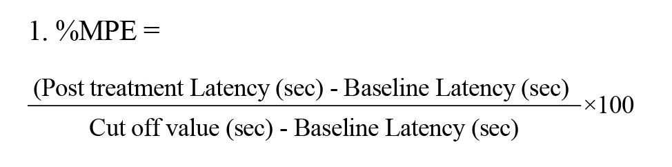
The tail-flick test was performed according to the method described by D’Amour and Smith (1941). The Tail-flick device (Harvard, USA) is designed to accurately measure the pain threshold to a focused light stimulus on the rat’s tail. Heat is applied sequentially to a spot 3, 5, or 7 cm from the tip of the rat’s tail. The light intensity is manually adjusted to 45% of maximum intensity, which in intact rats produces a TFL in the range of 3 to 4 seconds. When the animal feels pain and flicks its tail, a sensor detects this, stops the timer, and turns off the lamp. Thus, the animal’s reaction time is automatically recorded (Tzschentke et al., 2007). It should be noted that a small box is used during the test instead of a restraint to avoid immobilization stress. Therefore, the rat is gently held in the corner of the box and its tail is fixed on the groove of the tail-flick device. The cut-off time is set to 10 seconds if the animal does not show any reaction to the heat applied to the tail, which results in high analgesia. For each animal, the TEL was measured twice before drug administration, at 60-second intervals from two different points at the end of the tail, and the average was calculated as the baseline latency. The tail-flick test was performed at intervals of 60, 45, 30, 15, and 5 minutes after the application of the limiting stress. The increase in the TEL was defined as analgesia, and to examine the effect of drugs, stress, and their interaction, the percentage of maximal possible effect (%MPE) was calculated according to the Equation 1 (Reisi et al., 2014):
Experimental design
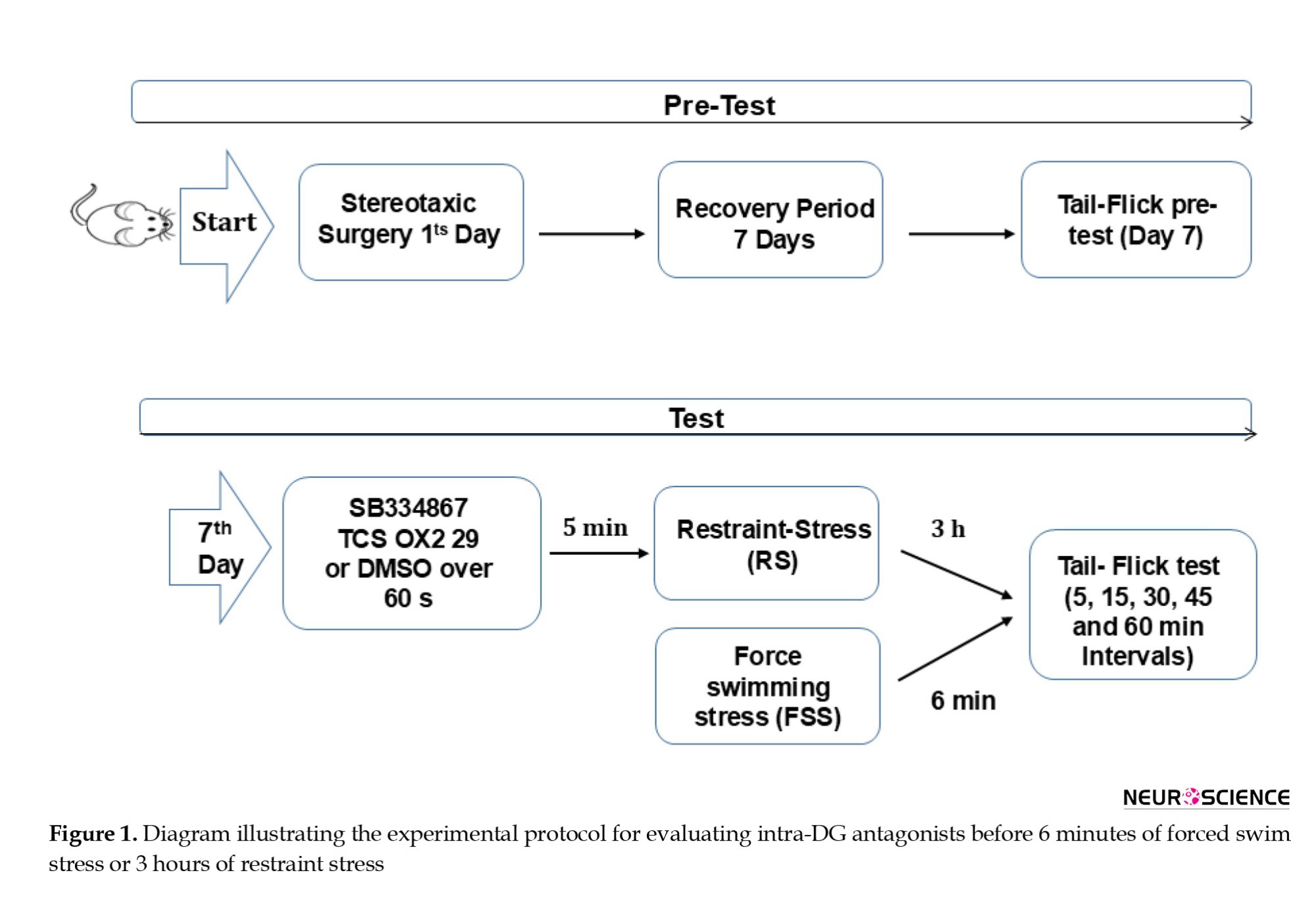
The present study was divided into two main experiments (Figure 1).
Pain is a multifaceted phenomenon and is classified into sensory, emotional, and cognitive components that are influenced by different neurobiological mechanisms (Khera & Rangasamy, 2021). Pain is also one of the somatic sensory modalities that has an important protective function and serves as a warning when tissue damage occurs or is likely to occur in order to prevent it. Three stages of pain have been proposed. The acute stage is caused by an acute and short-term painful stimulus. The second stage or chronic stage occurs when the stimulus is long-term and is accompanied by inflammation and causes tissue damage. The third stage, called neuropathic pain, is associated with peripheral or central nerve damage. Aδ and C afferent fibers transmit acute and chronic pain, respectively (Danziger, 2008).
Various studies have shown that stress may induce multiple changes in neural and hormonal status and induce analgesia in animals and humans. This phenomenon is defined as stress-induced analgesia (SIA) and is activated by endogenous pain inhibitory systems (Ahmad & Zakaria, 2015; Ford & Finn, 2008). Acute stress exposure can activate dopamine neurons through the release of substance P and endogenous opioids (Bannon et al., 1983). Pain influences stress behaviors, and stress modulates pain responses. Depending on the activities performed, various stressors are able to inhibit the response to noxious stimuli and lead to analgesia, including the restraint and forced-swim models (D’amour & Smith, 1941). Immobilization stress has often been used as a stimulus to study the effect of stress on pain in laboratory mice and rats (Cruthirds et al., 2011). In addition, the tail-flick test, hot plate test, and formalin test are commonly used as a noxious stimulus after exposure to stress paradigms (Malmberg & Bannon, 1999). Studies have shown that forced swimming stress increases the jump latency in the hot plate test and the tail-flick latency (TFL) (Ibironke & Rasak, 2013).
A large number of neurotransmitters and neuropeptides are involved in inhibiting and modulating the sensation of pain. Among these, endogenous opioid systems, dopamine, monoamines (serotonin and norepinephrine), endogenous cannabinoids, and gamma-aminobutyric acid can be mentioned (Korotkova et al., 2003; Liu & Song, 2025). In particular, the mesolimbic dopamine system is important in modulating pain and producing analgesia, and is activated under stress. Stress can induce analgesia through the release of neurotransmitters and neuropeptides. Studies have shown that the dopaminergic system and dopamine play a key role in endogenous analgesia and modulating pain in several areas of the central nervous system (CNS) (Li et al., 2019; Wood, 2008). One of the brain areas in which dopamine plays an active role is the mesolimbic system (Ikemoto, 2010). The mesolimbic system is a CNS circuit in which dopaminergic inputs from the ventral tegmental area innervate brain areas involved in executive, emotional, and motivational functions, including the prefrontal cortex (PFC), amygdala, and nucleus accumbens (Alcaro et al., 2007). Five dopamine receptor subtypes have been defined using molecular simulation methods, with D1 and D5 subtypes belonging to the D1 subfamily, while D2, D3, and D4 belong to the D2 subfamily of dopamine receptors. Dopamine receptor subtypes have different affinities for different ligands (Martel & Gatti McArthur, 2020). Acute stress exposure has been shown to increase extracellular dopamine levels in the nucleus accumbens and related regions (Vaessen et al., 2015).
The hippocampal formation (HF) is a C-shaped structure in the floor of the temporal horn of the lateral ventricle, recognized as a prominent component of the limbic system. It was named by Arantius (1587), who derived the word from the Greek word for seahorse (Adedayo et al., 2023; Insausti & Amaral, 2004). According to Insausti and Amaral (2004), the HF is composed of six distinct structures, including the entorhinal cortex, parasubiculum, perisubiculum, subiculum major (CA1-CA3), hippocampus, and dentate gyrus (DG) (Insausti & Amaral, 2004). The DG is described as the prominent gateway of the HF that conveys cortical afferents to the limbic system, among other components of the HF (Scharfman, 2011). Furthermore, it has been shown that the DG as a part of the HF plays an undeniable role in the processing of pain-related information (Boltyansky, 2018) through various mechanisms and receptors within the DG, such as N-methyl-D-aspartate (NMDA) receptors (McKenna & Melzack, 2001), histamine receptors (Khalilzadeh et al., 2010) and orexin receptors (Bolouri-Roudsari et al., 2024). Also, chemical stimulation of the lateral hypothalamus (LH) can induce analgesia through the involvement of dopaminergic receptors in the DG (Khaleghzadeh-Ahangar et al., 2021).
Studies have shown that dopamine release blocks pain in the mesolimbic/mesocortical pathway, and this effect is reduced by lesioning dopaminergic neurons in the ventral tegmental area (Wood, 2006). In 1990, Morgan and Franklin showed that depletion of dopamine in the ventral tegmental area (VTA) using 6-hydroxydopamine (6-OHDA) reduced the analgesic effects of systemically administered morphine and diamphetamine in the formalin model of inflammatory pain (Morgan & Franklin, 1990). A recent study showed that forced swim stress exposure can significantly increase analgesic responses in an acute pain model (Noursadeghi et al., 2022). On the other hand, FSS-induced analgesia was attenuated by blockade of D2-like dopamine receptors in the DG in an inflammatory pain model (Merdasi et al., 2022). Although considerable evidence suggests that the DG is involved in processing pain-related information and that dopaminergic receptors are involved in SIA, there is no study comparing the effects of different types of stress on dopamine receptor-mediated pain reduction in the tail-flick test.
A recent study has shown that dopamine receptor subtypes play an important role in behavioral and neurochemical adaptations to stress. Evidence suggests that D1 receptor activation is associated with the regulation of prefrontal and limbic circuitry in stress, particularly in sustained psychological stressors, such as restraint stress, which is associated with prolonged increases in glucocorticoids and altered dopamine turnover (Nazari-Serenjeh et al., 2024). In contrast, D2 receptors have been more closely associated with coping strategies and motivational components of the stress response, particularly in paradigms that simultaneously involve psychological and physical stressors, such as forced swim stress, where differential receptor engagement has been reported (Noursadeghi et al., 2022). This difference in receptor engagement pattern is likely due to differences in downstream signaling pathways and their specific regional distribution in the brain. Therefore, this study aimed to comparatively investigate the role of D1 and D2 receptors within the DG in two types of RS and FSS in the acute pain.
Materials and Methods
Animals
One hundred and thirty-three male Wistar albino rats weighing 220–250 g were used in this study. All animals were purchased from the Pasteur Institute, Tehran, Iran, and were maintained at standard temperature and humidity (23±1 °C, 50±5% relative humidity) under a 12-h light/dark cycle (lights on from 7 am to 7 pm). Adequate food and water were available ad libitum. All procedures were performed in accordance with the guidelines outlined in the National Institutes of Health Guide for the Care and Use of Laboratory Animals (NIH Publications; 8th edition, revised 2011), and were approved by the Research and Ethics Committee of Shahid Beheshti University of Medical Sciences.
Drugs
SCH-23390 and Sulpiride (Tocris Bioscience, Bristol, UK), as D1- and D2-like dopamine receptor antagonists (0.25, 1, and 4 μg/0.5 μL saline and DMSO, respectively, per rat), were dissolved in normal saline and 12% DMSO, respectively. The drugs were freshly prepared on the test day and microinjected into the DG. The concentration of the drugs was chosen based on previous reports of animals’ behavior (Matini et al., 2020; Siahposht-Khachaki et al., 2021).
Stereotaxic surgery
Approximately 5–10 min before stereotaxic surgery, subjects were injected intraperitoneally with a mixture of 10% ketamine (100 mg/kg) and 2% xylazine (10 mg/kg) and were deeply anesthetized after 3–5 min. The rats’ scalps were then shaved, and subjects were placed in a stereotaxic apparatus (Stolting, Wooddale, IL, USA). Subsequently, a lidocaine/epinephrine mixture (0.2 mL) was injected subcutaneously around the surgical site to minimize bleeding and pain responses. Bregma and lambda were located after scalp removal, and DG coordinates were calculated following the Paxinos and Watson rat brain atlas (Paxinos and Watson, 2006) (AP=3.7±2 mm posterior to bregma, LM=±2.2 mm lateral to midline, and DV=3.6 mm ventral to the skull surface). A 6 mm guide cannula (23-gauge stainless steel) was implanted unilaterally, 1 mm superior to the hippocampal DG, to the left or right of the midline of each rat randomly, and was fixed to the skull surface by two stainless steel screws and dental acrylic cement. After 24 hours, the rats were housed 2–3 per cage and given a median recovery period of 6 days.
Intra-DG administration
Different doses of D1- and D2-like antagonists (0.25, 1, and 4 μg/0.5 μL) or vehicle (Saline and 12% DMSO) were administered intra-DG. The injection volume was 0.5 μL, performed using a 1 μL Hamilton syringe. The microinjection process took approximately 60 seconds, and the needle was held in place for an additional 60 seconds to optimize drug release and minimize backflow.
Stress induction
Forced swim stress
For acclimatization, subjects were transferred to the testing room half an hour before the start of the experiments. Five minutes after the injection of the drug into the DG or microinjection of the vehicle, subjects were placed in a Plexiglas tank with a diameter of 30 cm and a height of 50 cm, which was filled to more than 30 cm with clean water (25±1 °C) for 6 minutes. After that, subjects were removed from the tank and gently dried with a towel (Askari et al., 2021; Saghafi et al., 2025).
Restraint stress
Five minutes after the administration of antagonist or vehicle, to induce acute immobilization stress, rats were placed in a 25 cm long and 6 cm wide Plexiglas tubular restraint for 3 hours, the length of which was adjustable to prevent the animal from moving. After each test, they were placed in their cage for at least 10 minutes before subsequent tests.
Tail-flick test
The tail-flick test was performed according to the method described by D’Amour and Smith (1941). The Tail-flick device (Harvard, USA) is designed to accurately measure the pain threshold to a focused light stimulus on the rat’s tail. Heat is applied sequentially to a spot 3, 5, or 7 cm from the tip of the rat’s tail. The light intensity is manually adjusted to 45% of maximum intensity, which in intact rats produces a TFL in the range of 3 to 4 seconds. When the animal feels pain and flicks its tail, a sensor detects this, stops the timer, and turns off the lamp. Thus, the animal’s reaction time is automatically recorded (Tzschentke et al., 2007). It should be noted that a small box is used during the test instead of a restraint to avoid immobilization stress. Therefore, the rat is gently held in the corner of the box and its tail is fixed on the groove of the tail-flick device. The cut-off time is set to 10 seconds if the animal does not show any reaction to the heat applied to the tail, which results in high analgesia. For each animal, the TEL was measured twice before drug administration, at 60-second intervals from two different points at the end of the tail, and the average was calculated as the baseline latency. The tail-flick test was performed at intervals of 60, 45, 30, 15, and 5 minutes after the application of the limiting stress. The increase in the TEL was defined as analgesia, and to examine the effect of drugs, stress, and their interaction, the percentage of maximal possible effect (%MPE) was calculated according to the Equation 1 (Reisi et al., 2014):
Experimental design
The present study was divided into two main experiments (Figure 1).
The first experiment was considered to investigate the possible effects of cannulation or microinjection volume on analgesia induced by FSS or RS. Animals in this group were divided into six control subgroups (n=6-8 animals per group):
a) Intact subgroup (no vehicle injection + no stress): animals underwent the tail-flick test only.
b) Sham subgroup (vehicle injection + no stress): animals cannulated in the DG underwent the tail-flick test only.
c) Forced swim stress (FSS) control subgroup (no vehicle + FSS): animals were exposed to FSS for 6 minutes between the baseline and post-stress delay phases.
d) RS control subgroup (no vehicle + RS): animals were exposed to RS for 3 hours between the baseline and post-stress delay phases.
e) Control subgroup (vehicle + FSS or RS): animals received both 5 μL of vehicle and 6 minutes of FSS or 3 hours of RS between the baseline delay phase and the delay phase after drug administration.
The second experiment involved groups receiving SCH23390 or sulpiride and was designed to investigate the effect of intra-DG administration of dopamine receptor antagonists on analgesia induced by FSS or RS. In this set of experiments, rats were divided into eight distinct subgroups. In order to assess the role of dopamine receptors in the DG in analgesic responses induced by FSS or RS, different doses of SCH23390 or sulpiride (0.25, 1, and 4 μg in a volume of 0.5 μL) were injected unilaterally into the DG just 5 min before stress exposure. The vehicle group (saline; 0.5 μL + FSS or RS) also received 0.5 μL of normal saline intra-DG as a drug vehicle 5 min before FSS or RS.
Histological confirmation
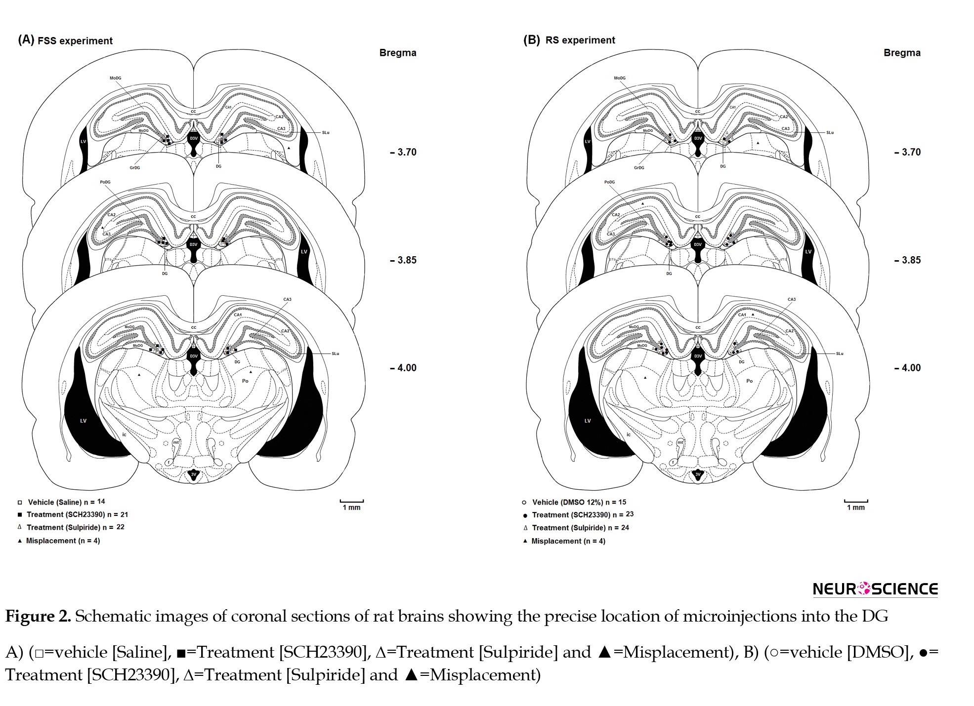
At the end of the experiments, the animals were completely anesthetized by intraperitoneal injection of ketamine and xylazine. They were then sacrificed and their brains were removed and fixed in 10% formalin solution. After 4 days, the brains were cut into 50 μm sections using a vibratome and these sections were examined under a stereomicroscope. The location of the guide cannula tip was compared with the corresponding sections in a rat brain atlas (Paxinos & Watson, 2004), (Figure 2).
a) Intact subgroup (no vehicle injection + no stress): animals underwent the tail-flick test only.
b) Sham subgroup (vehicle injection + no stress): animals cannulated in the DG underwent the tail-flick test only.
c) Forced swim stress (FSS) control subgroup (no vehicle + FSS): animals were exposed to FSS for 6 minutes between the baseline and post-stress delay phases.
d) RS control subgroup (no vehicle + RS): animals were exposed to RS for 3 hours between the baseline and post-stress delay phases.
e) Control subgroup (vehicle + FSS or RS): animals received both 5 μL of vehicle and 6 minutes of FSS or 3 hours of RS between the baseline delay phase and the delay phase after drug administration.
The second experiment involved groups receiving SCH23390 or sulpiride and was designed to investigate the effect of intra-DG administration of dopamine receptor antagonists on analgesia induced by FSS or RS. In this set of experiments, rats were divided into eight distinct subgroups. In order to assess the role of dopamine receptors in the DG in analgesic responses induced by FSS or RS, different doses of SCH23390 or sulpiride (0.25, 1, and 4 μg in a volume of 0.5 μL) were injected unilaterally into the DG just 5 min before stress exposure. The vehicle group (saline; 0.5 μL + FSS or RS) also received 0.5 μL of normal saline intra-DG as a drug vehicle 5 min before FSS or RS.
Histological confirmation
At the end of the experiments, the animals were completely anesthetized by intraperitoneal injection of ketamine and xylazine. They were then sacrificed and their brains were removed and fixed in 10% formalin solution. After 4 days, the brains were cut into 50 μm sections using a vibratome and these sections were examined under a stereomicroscope. The location of the guide cannula tip was compared with the corresponding sections in a rat brain atlas (Paxinos & Watson, 2004), (Figure 2).
Locomotor activity
The total distance traveled by the subject in an open field (60×60 cm2) over 60 min was tracked using a 3-CCD camera (Panasonic Corporation, Osaka, Japan), and the data were analyzed by Ethovision software (version 7; Noldus Information Technology, Wageningen, The Netherlands).
Statistical analysis
Statistical analysis of data was performed using GraphPad Prism software, version 6 (GraphPad Software, San Diego, USA). Results were reported as Mean±SEM, and normality of data distribution was assessed using the Kolmogorov–Smirnov test. Two-way repeated measures ANOVA with Bonferroni post hoc test was used to compare the percentage of maximum possible effect at different times between treatment groups. Also, one-way ANOVA followed by Tukey’s post hoc test was used to compare groups, when necessary.
To evaluate the effect of RS and FSS on acute pain behaviors, the area under the curve (AUC) values of different groups were calculated, and the unpaired t-test was used to compare the groups. In all analyses, a statistical significance level of P<0.05 was considered.
Results
Effects of RS and FSS on antinociceptive behaviors in the tail-flick test
Pain-related behavior in the tail-flick test, as an animal model of acute pain, was assessed at time intervals of 5, 15, 30, 45, and 60 minutes, and the MPE% was obtained, and the AUC of the data related to TFL times after the application of the painful stimulus was examined. Two-way analysis of variance (ANOVA) followed by Bonferroni post-test showed that pain responses in the tail-flick test were significantly reduced in the groups exposed to RS and FSS compared to the non-stress groups (Figure 3A) [treatment effect: F5, 180=108.3, P<0.0001; time effect: F4, 180=3.731, P=0.006; treatment and time interaction effect: F20, 180=1.090, P=0.3629].
In addition, to evaluate the effect of RS and FSS on acute pain behaviors, the AUC values of different groups were calculated and one-way ANOVA was used to compare the groups. The AUC values in the NO DMSO + RS group increased significantly compared to the No DMSO + No RS group (P<0.0001). Similarly, a significant increase was observed in the DMSO + RS group compared to the DMSO + No RS group (P<0.0001). Similarly, the AUC values in the No DMSO + FSS group increased significantly compared to the No DMSO + No FSS group (P<0.0001). Also, a significant increase was observed in the DMSO + FSS group compared to the DMSO + No RS group (P<0.0001) (Figure 3B).
Effect of intra-DG administration of SCH-23390 on the antinociceptive behavior produced by RS and FSS
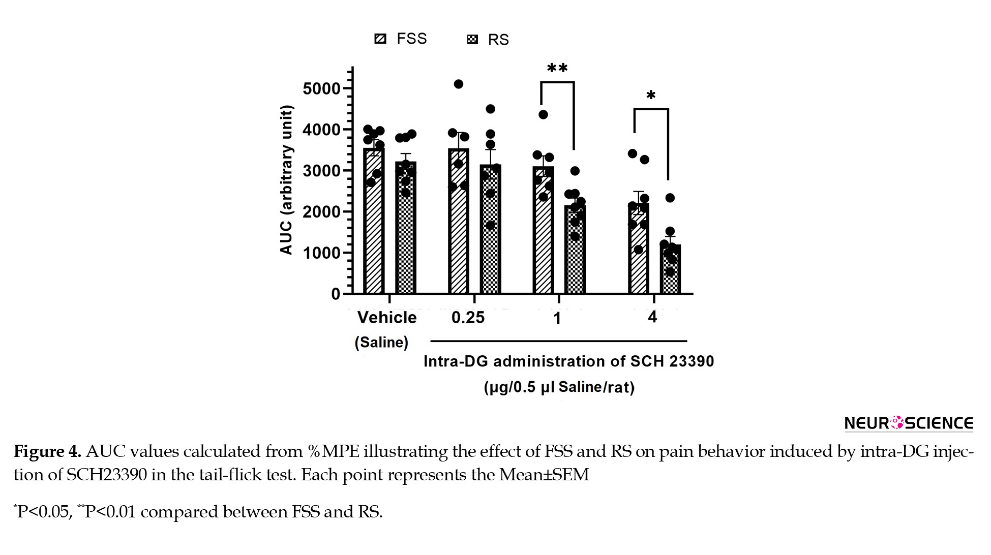
According to the results of two-way ANOVA followed by Bonferroni post hoc test, there was a significant difference between the RS and FSS groups in pain behaviors induced by microinjection of SCH-23390 (0.25, 1 and 4 μg/0.5 μL) into the DG in the tail flick test (Figure 4) (stress effect: F1, 51=13.67, P<0.005; treatment effect: F3, 51=19.7, P<0.0001; stress and treatment interaction effect: F3, 51=0.99, P=0.404).
Effect of intra-DG administration of SCH-23390 on the antinociceptive behavior produced by RS and FSS
According to the results of two-way ANOVA followed by Bonferroni post hoc test, there was a significant difference between the RS and FSS groups in pain behaviors induced by microinjection of SCH-23390 (0.25, 1 and 4 μg/0.5 μL) into the DG in the tail flick test (Figure 4) (stress effect: F1, 51=13.67, P<0.005; treatment effect: F3, 51=19.7, P<0.0001; stress and treatment interaction effect: F3, 51=0.99, P=0.404).
Also, to evaluate the effect of RS and FSS on acute pain behaviors, the AUC values of different groups were calculated and the unpaired t-test was used to compare the groups. There was a significant difference between RS and FSS in the SCH-23390 groups (1 µg/0.5 μL, P<0.01 and 4 µg/0.5 μL, P<0.05). The results of this experiment showed that intra-DG administration of SCH-23390 significantly reduced the analgesic responses induced by RS more than those induced by FSS, indicating a greater involvement of D1 receptor signaling in RS-induced analgesia.
Effect of intra-DG administration of sulpiride on the antinociceptive behavior produced by RS and FSS
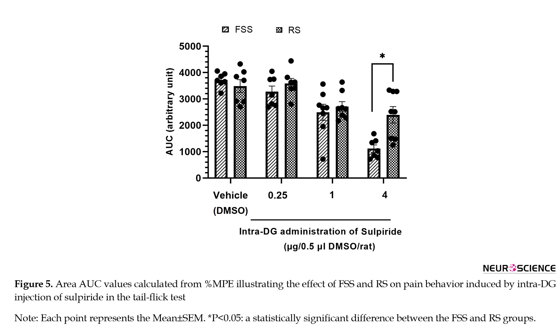
Two-way ANOVA followed by Bonferroni post hoc test showed that the effects of RS and FSS on pain behaviors induced by microinjection of sulpiride (0.25, 1, and 4 μg/0.5 μL) into the DG in the tail flick test were significant (Figure 5) (stress effect: F1, 51=6.009, P=0.017; treatment effect: F3, 51=27.48, P<0.0001; stress and treatment interaction effect: F3, 51=3.865, P=0.014).
Effect of intra-DG administration of sulpiride on the antinociceptive behavior produced by RS and FSS
Two-way ANOVA followed by Bonferroni post hoc test showed that the effects of RS and FSS on pain behaviors induced by microinjection of sulpiride (0.25, 1, and 4 μg/0.5 μL) into the DG in the tail flick test were significant (Figure 5) (stress effect: F1, 51=6.009, P=0.017; treatment effect: F3, 51=27.48, P<0.0001; stress and treatment interaction effect: F3, 51=3.865, P=0.014).
In addition, to evaluate the effect of RS and FSS on acute pain behaviors, the AUC values of different groups were calculated and the unpaired t-test was used to compare the groups. There was a significant difference between RS and FSS in the sulpiride (4 µg/0.5 μL) groups (P<0.05). These findings suggest that signaling through D2 receptors plays a more prominent role in eliciting analgesic responses during FSS compared to RS.
Effect of intra-DG microinjection of dopamine receptor antagonists on locomotor activity
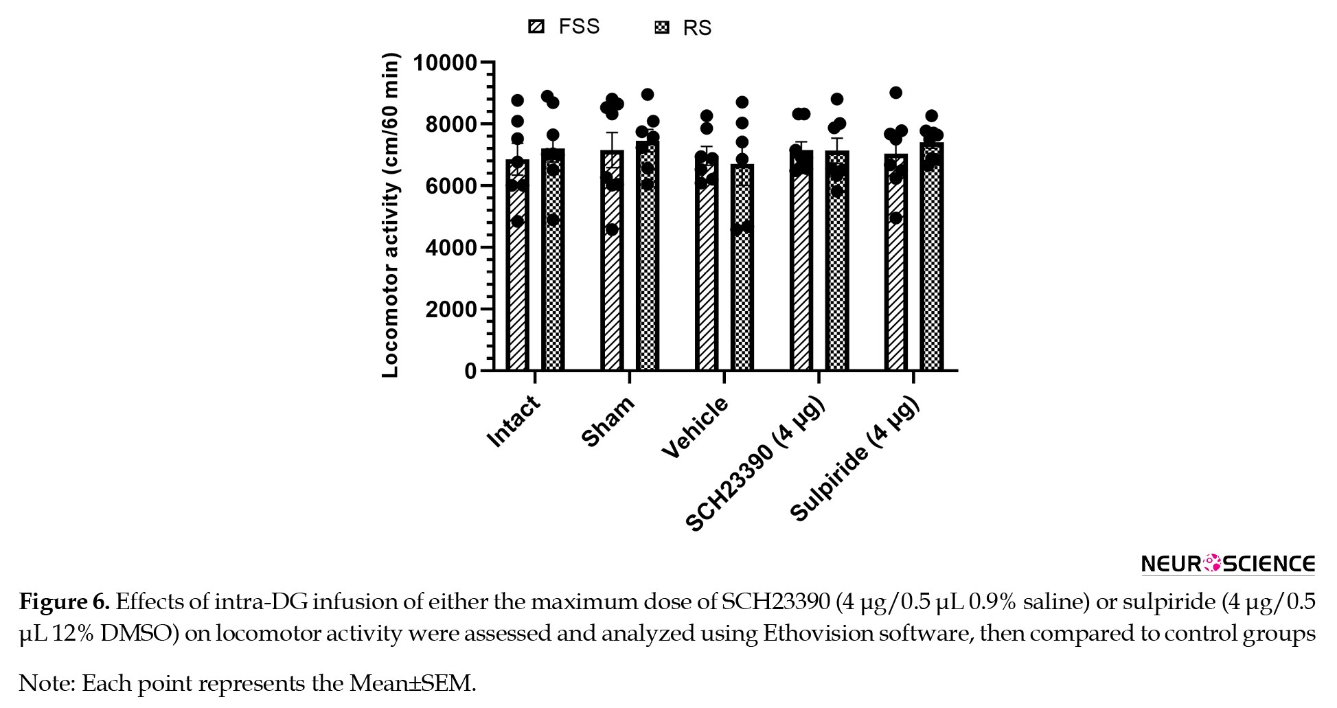
The traveled distance was measured over 60 min as an indicator of locomotor activity. One-way ANOVA followed by Tukey’s multiple comparison test showed that none of the experimental procedures affected locomotor activity (stress effect: F1, 64 =0.2990, P=0.5864; treatment effect: F4, 64=0.3285, P=0.8578; stress and treatment interaction effect: F4, 64=0.1944, P=0.9405; [Figure 6]).
Effect of intra-DG microinjection of dopamine receptor antagonists on locomotor activity
The traveled distance was measured over 60 min as an indicator of locomotor activity. One-way ANOVA followed by Tukey’s multiple comparison test showed that none of the experimental procedures affected locomotor activity (stress effect: F1, 64 =0.2990, P=0.5864; treatment effect: F4, 64=0.3285, P=0.8578; stress and treatment interaction effect: F4, 64=0.1944, P=0.9405; [Figure 6]).
Discussion
The most important findings of the present investigation were as follows: (a) RS and FSS significantly increased antinociceptive behaviors in an acute model of pain compared to the control group; (b) intra-DG administration of SCH-23390 significantly reduced antinociceptive behaviors in the RS group compared to the FSS group; (c) the effect of FSS was more remarkable than that of RS on antinociceptive behaviors in groups that received sulpiride in the DG.
The DG, as part of the hippocampus, plays a key role in emotional processing, learning, and memory (Sun et al., 2023; Tyng et al., 2017). Kesner showed that the DG plays a fundamental role in relaying cortical inputs to the limbic system and in integrating emotional responses (Kesner, 2007). Since both pain and stress are associated with emotional processing, changes in dopamine receptor activity in the DG could directly influence the coordination of behavioral and neurochemical responses to stress.
The phenomenon of pain inhibition in response to stress is known as SIA (al’Absi et al., 2021). Exposure to stressful or fearful stimuli, whether conditioned or unconditioned, serves as a template for activating the endogenous pain inhibition system and can elicit this response (Taylor, 2017). The function of SIA is influenced by descending pain inhibition pathways, and any change in the activity of these pathways can disrupt it (Kwon et al., 2014). This suggests a complex and bidirectional interaction between stress and pain, in that pain can lead to stress, and stress can, depending on the circumstances, reduce or increase the intensity of pain (Melzack, 1999). The type of stressor determines the neural circuitry involved in pain inhibition. In general, mild stressors act primarily through mechanisms dependent on the endogenous opioid system, whereas severe stressors activate more opioid-independent pathways (Ferdousi & Finn, 2018). Furthermore, evidence suggests that different types of stressors can exert multifaceted and complex effects on dopamine levels in the mesolimbic system (Baik, 2020).
Several studies have shown that various forms of stress, including forced swimming, restraint, sleep deprivation, and food restriction or deprivation, can alter the threshold and intensity of pain perception (Butler & Finn, 2009). In the present study, RS and FSS were used as aversive stressors to assess the effect of stress on pain responses in rats. The findings showed that RS and FSS administration resulted in an analgesic effect in acute pain and significantly increased the latency to respond during a 60-min tail-flick test. The differential effect of sulpiride in various stress paradigms suggests that different neurochemical mechanisms are responsible for SIA, and this depends on the type of stressor. While D1 receptor signaling is dominant in RS, D2 pathways may be more activated in FSS, suggesting a specific involvement of dopaminergic circuits depending on the type of stressor. These results are consistent with previous reports that have confirmed the inhibitory effect of stress in animal models of thermal and visceral pain (Askari et al., 2021; Faramarzi et al., 2016).
SIA plays an important role in the temporary inhibition of nociceptive transmission as an adaptive mechanism in the face of environmental stressors and has attracted the attention of researchers to identify the central neural networks involved in pain inhibition (Butler & Finn, 2009). Although this phenomenon was initially thought to be mediated mainly by the endogenous opioid system, subsequent evidence has shown that non-opioid mechanisms also contribute significantly (Mogil et al., 1996). For example, in the dorsal hippocampus of the guinea pig, pain transmission has been shown to be regulated by the interaction of GABAergic, cholinergic, and opioidergic systems (Mendes & Menescal-de-Oliveira, 2008). Furthermore, during SIA, activation of a network of neurotransmitters and neuromodulators, including monoamines, GABA, glutamate, and endocannabinoids has been reported, indicating the multisystem and complex nature of this phenomenon (Butler & Finn, 2009).
Our results indicated that intra-DG administration of SCH-23390 significantly reduced antinociceptive behaviors in RS more than in FSS, but the effect of FSS was more remarkable than RS on antinociceptive behaviors in groups that received sulpiride in the DG.
The findings suggest that individual susceptibility to SIA is highly variable and is associated with a variety of characteristics, including the type and intensity of stress and the degree of opioid sensitivity and response to shocking stimuli. In addition, SIA is influenced by environmental and individual factors, such as age, gender, and previous painful or stressful experiences (Butler & Finn, 2009). In a recent study, the application of three hours of RS induced SIA in both the acute and inflammatory phases of the formalin test. However, when dopamine receptor antagonists were microinjected into the DG, the intensity of this analgesic response was significantly reduced in both phases of the test (Nazari-Serenjeh et al., 2024). These findings suggest that various kinds of stressors act in pain modulation in descending pathways through specific receptors. However, to more precisely elucidate the contribution of each of these receptor subtypes in the process of pain modulation, a comprehensive and integrated approach is necessary.
Evidence suggests that RS and FSS can induce different patterns of dopamine release in the DG and extended hippocampal circuits. RS, as a long-term psychological stimulus, often induces a sustained and moderate release of dopamine that activates mostly high-affinity D1 receptors. In contrast, FSS, which is short-term but highly physically demanding, can induce a rapid and phasic release of dopamine and more effectively activates D2 receptors, pathways that are associated with motivational and coping responses (Barrot et al., 2000, Kalivas & Duffy, 1995). RS primarily activates cortico-limbic afferents, including inputs from the PFC and amygdala (McKlveen et al., 2015), regions that are strongly modulated by D1 receptor-mediated dopaminergic signaling (Seamans & Yang, 2004). In contrast, FSS engages broader motivational and mesolimbic circuits, particularly the ventral tegmental area–nucleus accumbens pathway (Kalivas & Duffy, 1995; Russo & Nestler, 2013), where D2 receptor signaling plays a prominent regulatory role (Beaulieu & Gainetdinov, 2011). Such circuit-specific dopaminergic organization may explain the differential receptor contributions observed across stress paradigms (Grace, 2016).
The temporal and qualitative differences between RS (3 hours) and FSS (6 minutes) likely contribute to receptor specificity. Studies have shown that dopamine release dynamics differ depending on the duration and nature of stimuli — tonic, sustained dopamine signaling predominates during prolonged stimuli, whereas short, intense stimuli evoke rapid, phasic dopamine release patterns that shape receptor activation profiles (Grace, 1991; Baik, 2020). Prolonged RS may evoke sustained dopaminergic tone favoring D1 receptor-mediated modulation of analgesia, as prolonged exposure to psychological stressors has been associated with persistent dopaminergic activity that influences receptor responsiveness over longer time frames. Sustained dopaminergic tone is linked to differential engagement of postsynaptic receptors, which may bias toward D1-mediated signaling under longer stress exposures, whereas the brief, high-intensity FSS may produce rapid, transient dopamine surges more suited to D2 receptor activation. Acute and intense stressors produce burst firing and rapid dopamine efflux, which has been shown to differentially influence downstream receptor activity, often invoking phasic signaling that may more potently activate receptor subtypes, including D2 with high temporal sensitivity. These differences highlight how stressor intensity and duration shape dopaminergic involvement in analgesic responses. Research supports the idea that stressor intensity, controllability, and duration affect both tonic and phasic dopamine release, thereby modulating how dopamine contributes to behavioral and analgesic outcomes.
Anatomical and molecular studies demonstrate that dopamine receptor subtypes show heterogeneous distribution across hippocampal subfields, including the DG (Gangarossa et al., 2012). D1 receptors are primarily localized to granule cells in the dorsal DG, supporting cognitive-emotional integration. Evidence indicates that D1 receptors are prominently expressed in granule neurons of the DG and play a key role in hippocampal-dependent cognitive processing and synaptic plasticity (Gangarossa et al., 2012; Hansen & Manahan-Vaughan, 2014), while D2 receptors are enriched in ventral DG circuits associated with motivational and coping behaviors. The ventral hippocampus, including ventral DG circuits, is more strongly connected to limbic and motivational systems, and D2 receptor signaling has been implicated in emotional and motivational regulation within these pathways (Beaulieu & Gainetdinov, 2011; Fanselow & Dong, 2010). Such localization may explain the stressor-specific recruitment of receptor subtypes and aligns with the observed differential effects of SCH-23390 and sulpiride. Functional differentiation between dorsal (cognitive) and ventral (affective–motivational) hippocampal circuits supports the idea that receptor localization contributes to stressor-specific behavioral and analgesic outcomes (Fanselow & Dong, 2010; Baik, 2020).
In summary, the present study demonstrates that stressor type selectively engages dopaminergic receptor subtypes in the DG, with RS predominantly recruiting D1 receptor-mediated pathways and FSS preferentially engaging D2 receptor-dependent mechanisms. This finding provides novel mechanistic insight into how different forms of stress modulate analgesic responses and highlights the importance of stressor-specific dopaminergic signaling in pain regulation. These results advance our understanding of the neurochemical underpinnings of SIA and provide a foundation for future research targeting receptor-specific interventions for stress- and pain-related disorders. The simultaneous use of electrophysiological techniques to assess the functional activity of neurons, molecular methods to measure gene and protein expression, cellular studies to investigate intracellular mechanisms, and immunohistochemical analyses to determine the location and distribution pattern of receptors can provide a more accurate picture of the role of these receptors in the regulation of pain pathways.
Ethical Considerations
Compliance with ethical guidelines
This study was approved by the Research Ethics Committee of Shahid Beheshti University of Medical Sciences, Tehran, Iran (Code: IR.SBMU.AEC.1404.022).
Funding
This study was extracted from the PhD dissertation of Homayoon Golmohammadi, approved by the Department of Physiology and Pharmacology, Faculty of Medical Sciences, Shahid Beheshti University of Medical Sciences, Tehran, Iran. This dissertation was supported by the Vice-Chancellor for Research & Technology of Shahid Beheshti University of Medical Sciences (Grant No.: 03-43013612-1404/02/21). This project was also supported by Elite Researcher Grant Committee from the National Institute for Medical Research Development (NIMAD), Tehran, Iran (Award No.: 4030151).
Authors' contributions
Conceptualization and study design: Abbas Haghparast and Mohadeseh Ghalandari-Shamami; Data acquisition: Homayoon Golmohammadi; Data analysis and interpretation: Abbas Haghparast; Writing the original draft: Mohadeseh Ghalandari-Shamami; Review and editing: Abbas Haghparast and Mohadeseh Ghalandari-Shamami; Final approval: All authors.
Conflict of interest
The authors declared no conflict of interest.
Acknowledgments
The authors would like to thank the Neuroscience Research Center, Institute of Neuroscience and Cognition, Shahid Beheshti University of Medical Sciences, Tehran, Iran for its cooperation in conducting this study.
References
Adedayo, L., Ojo, G., Umanah, S., Aitokhuehi, G., Emmanuel, I.-O., & Bamidele, O. (2023). Hippocampus: Its Role in Relational Memory. In Hippocampus - More than Just Memory. London: IntechOpen. [DOI:10.5772/intechopen.111478]
Ahmad, A. H., & Zakaria, R. (2015). Pain in times of stress. The Malaysian Journal of Medical Sciences: MJMS, 22(Spec Issue), 52-61. [PMID]
al’Absi, M., Nakajima, M., & Bruehl, S. (2021). Stress and pain: Modality-specific opioid mediation of stress-induced analgesia. Journal of Neural Transmission, 128(9), 1397-1407. [DOI:10.1007/s00702-021-02401-4] [PMID]
Alcaro, A., Huber, R., & Panksepp, J. (2007). Behavioral functions of the mesolimbic dopaminergic system: An affective neuroethological perspective. Brain Research Reviews, 56(2), 283-321. [DOI:10.1016/j.brainresrev.2007.07.014] [PMID]
Askari, K., Oryan, S., Eidi, A., Zaringhalam, J., & Haghparast, A. (2021). Modulatory role of the orexin system in stress‐induced analgesia: Involvement of the ventral tegmental area. European Journal of Pain, 25(10), 2266-2277. [DOI:10.1002/ejp.1840] [PMID]
Baik, J. H. (2020). Stress and the dopaminergic reward system. Experimental & Molecular Medicine, 52(12), 1879-1890. [DOI:10.1038/s12276-020-00532-4] [PMID]
Bannon, M. J., Elliott, P. J., Alpert, J. E., Goedert, M., Iversen, S. D., & Iversen, L. L. (1983). Role of endogenous substance P in stress-induced activation of mesocortical dopamine neurones. Nature, 306(5945), 791–792. [DOI:10.1038/306791a0] [PMID]
Barrot, M., Marinelli, M., Abrous, D. N., Rougé-Pont, F., Le Moal, M., & Piazza, P. V. (2000). The dopaminergic hyper-responsiveness of the shell of the nucleus accumbens is hormone-dependent. The European Journal of Neuroscience, 12(3), 973–979. [DOI:10.1046/j.1460-9568.2000.00996.x] [PMID]
Beaulieu, J. M., & Gainetdinov, R. R. (2011). The physiology, signaling, and pharmacology of dopamine receptors. Pharmacological Reviews, 63(1), 182-217. [DOI:10.1124/pr.110.002642] [PMID]
Bolouri-Roudsari, A., Baghani, M., Askari, K., Mazaheri, S., & Haghparast, A. (2024). The integrative role of orexin-1 and orexin-2 receptors within the hippocampal dentate gyrus in the modulation of the stress-induced antinociception in the formalin pain test in the rat. Behavioural Pharmacology, 35(1), 14-25. [DOI:10.1097/FBP.0000000000000737] [PMID]
Boltyansky, B. (2018). Novel aspects of neuropathic pain processing within emotion-associated higher brain regions: exemplified in the dentate gyrus [MSc thesis]. Haifa: University of Haifa. [Link]
Butler, R. K., & Finn, D. P. (2009). Stress-induced analgesia. Progress in Neurobiology, 88(3), 184-202. [DOI:10.1016/j.pneurobio.2009.04.003] [PMID]
Cruthirds, D. F., Siangco, A. L., Hartman, C. J., Sandefur, D. C., Spencer, J. M., & Dyer, C. A., et al. (2011). Effects of immobilization stress and hormonal treatment on nociception. AANA Journal, 79(5), 375–380. [PMID]
D’AMOUR, F. E., & SMITH, D. L. (1941). A method for determining loss of pain sensation. The Journal of Pharmacology and Experimental Therapeutics, 72(1), 74-79. [DOI:10.1016/S0022-3565(25)03823-6]
Danziger N. (2007). [Pathophysiology of pain in venous disease (French)]. Journal des Maladies Vasculaires, 32(1), 1–7. [DOI: 10.1016/j.jmv.2006.10.001] [PMID]
Faramarzi, G., Zendehdel, M., & Haghparast, A. (2016). D1‐and D2‐like dopamine receptors within the nucleus accumbens contribute to stress‐induced analgesia in formalin‐related pain behaviours in rats. European Journal of Pain, 20(9), 1423-1432. [DOI:10.1002/ejp.865] [PMID]
Fanselow, M. S., & Dong, H. W. (2010). Are the dorsal and ventral hippocampus functionally distinct structures? Neuron, 65(1), 7-19. [DOI:10.1016/j.neuron.2009.11.031] [PMID]
Ferdousi, M., & Finn, D. P. (2018). Stress-induced modulation of pain: role of the endogenous opioid system. Progress in Brain Research, 239, 121-177. [DOI:10.1016/bs.pbr.2018.07.002] [PMID]
Ford, G. K., & Finn, D. P. (2008). Clinical correlates of stress-induced analgesia: Evidence from pharmacological studies. Pain, 140(1), 3-7. [DOI:10.1016/j.pain.2008.09.023] [PMID]
Gangarossa, G., Longueville, S., De Bundel, D., Perroy, J., Hervé, D., & Girault, J. A., et al. (2012). Characterization of dopamine D1 and D2 receptor-expressing neurons in the mouse hippocampus. Hippocampus, 22(12), 2199–2207. [DOI:10.1002/hipo.22044] [PMID]
Grace, A. A. (2016). Dysregulation of the dopamine system in the pathophysiology of schizophrenia and depression. Nature Reviews. Neuroscience, 17(8), 524–532. [DOI:10.1038/nrn.2016.57] [PMID]
Hansen, N., & Manahan-Vaughan, D. (2014). Dopamine D1/D5 receptors mediate information storage in the hippocampus long-term plasticity. Cerebral Cortex (New York, N.Y.: 1991), 24(4), 845–858. [DOI:10.1093/cercor/bhs362] [PMID]
Ibironke, G., & Rasak, K. (2013). Forced swimming stress-related hypoalgesia: Nondependence on the histaminergic mechanisms. Neurophysiology, 45(4), 340-343. [DOI:10.1007/s11062-013-9378-1]
Ikemoto, S. (2010). Brain reward circuitry beyond the mesolimbic dopamine system: A neurobiological theory. Neuroscience & Biobehavioral Reviews, 35(2), 129-150. [DOI:10.1016/j.neubiorev.2010.02.001] [PMID]
Insausti, R., & Amaral, D. G. (2004). Hippocampal formation. In G. Paxinos & G. K. MAI (Eds.), The human nervous system (pp. 871-914). New York: Academic Press. [DOI:10.1016/B978-012547626-3/50024-7]
Kalivas, P. W., & Duffy, P. (1995). Selective activation of dopamine transmission in the shell of the nucleus accumbens by stress. Brain Research, 675(1-2), 325-328. [DOI:10.1016/0006-8993(95)00013-G] [PMID]
Kesner, R. P. (2007). A behavioral analysis of dentate gyrus function. Progress in Brain Research, 163, 567-576. [DOI:10.1016/s0079-6123(07)63030-1]
Khaleghzadeh-Ahangar, H., Rashvand, M., & Haghparast, A. (2021). Role of D1-and D2-like dopamine receptors within the dentate gyrus in antinociception induced by chemical stimulation of the lateral hypothalamus in an animal model of acute pain. Physiology & Behavior, 229, 113214. [DOI:10.1016/j.physbeh.2020.113214] [PMID]
Khalilzadeh, E., Tamaddonfard, E., Farshid, A. A., & Erfanparast, A. (2010). Microinjection of histamine into the dentate gyrus produces antinociception in the formalin test in rats. Pharmacology Biochemistry and Behavior, 97(2), 325-332. [DOI:10.1016/j.pbb.2010.08.018] [PMID]
Khera, T., & Rangasamy, V. (2021). Cognition and pain: A review. Frontiers in Psychology, 12, 673962. [DOI:10.3389/fpsyg.2021.673962] [PMID]
Korotkova, T. M., Sergeeva, O. A., Eriksson, K. S., Haas, H. L., & Brown, R. E. (2003). Excitation of ventral tegmental area dopaminergic and nondopaminergic neurons by orexins/hypocretins. The Journal of Neuroscience: The Official Journal of the Society for Neuroscience, 23(1), 7–11. [DOI:10.1523/JNEUROSCI.23-01-00007.2003] [PMID]
Kwon, M., Altin, M., Duenas, H., & Alev, L. (2014). The role of descending inhibitory pathways on chronic pain modulation and clinical implications. Pain Practice, 14(7), 656-667. [DOI:10.1111/papr.12145] [PMID]
Li, C., Liu, S., Lu, X., & Tao, F. (2019). Role of descending dopaminergic pathways in pain modulation. Current Neuropharmacology, 17(12), 1176-1182. [DOI:10.2174/1570159X17666190430102531] [PMID]
Liu, Z., & Song, S. (2025). Neurotransmitters and Receptors in Anesthesia and Pain Modulation. In Z. Liu & S. Song (Eds.), Neuroscience of Anesthesia: From Cellular Mechanisms to Clinical Applications (pp. 33-44). Cham: Springer. [DOI:10.1007/978-3-031-85156-8_4]
Malmberg, A. B., & Bannon, A. W. (1999). Models of nociception: Hot‐plate, tail‐flick, and formalin tests in rodents. Current Protocols in Neuroscience, 6(1), 8-9. [DOI:10.1002/0471142301.ns0809s06]
Martel, J. C., & Gatti McArthur, S. (2020). Dopamine receptor subtypes, physiology and pharmacology: New ligands and concepts in schizophrenia. Frontiers in Pharmacology, 11, 1003. [DOI:10.3389/fphar.2020.01003] [PMID]
Matini, T., Haghparast, A., Rezaee, L., Salehi, S., Tehranchi, A., & Haghparast, A. (2020). Role of Dopaminergic Receptors Within the Ventral Tegmental Area in Antinociception Induced by Chemical Stimulation of the Lateral Hypothalamus in an Animal Model of Orofacial Pain. Journal of Pain Research, 13, 1449–1460. [DOI:10.2147/jpr.S255250] [PMID]
McKenna, J. E., & Melzack, R. (2001). Blocking NMDA receptors in the hippocampal dentate gyrus with AP5 produces analgesia in the formalin pain test. Experimental Neurology, 172(1), 92-99. [DOI:10.1006/exnr.2001.7777] [PMID]
Melzack, R. (1999). Pain and stress: A new perspective. In R. J. Gatchel & D. C. Turk (Eds.), Psychosocial factors in pain: Critical perspectives (pp. 89-106). New York: Guilford Press. [Link]
Mendes, L. A. F., & Menescal-de-Oliveira, L. (2008). Role of cholinergic, opioidergic and GABAergic neurotransmission of the dorsal hippocampus in the modulation of nociception in guinea pigs. Life sciences, 83(19-20), 644-650. [DOI:10.1016/j.lfs.2008.09.006] [PMID]
Merdasi, P. G., Dezfouli, R. A., Mazaheri, S., & Haghparast, A. (2022). Blocking the dopaminergic receptors in the hippocampal dentate gyrus reduced the stress-induced analgesia in persistent inflammatory pain in the rat. Physiology & Behavior, 253, 113848. [DOI:10.1016/j.physbeh.2022.113848] [PMID]
McKlveen, J. M., Myers, B., & Herman, J. P. (2015). The medial prefrontal cortex: Coordinator of autonomic, neuroendocrine and behavioural responses to stress. Journal of Neuroendocrinology, 27(6), 446-456. [DOI:10.1111/jne.12272] [PMID]
Mogil, J. S., Sternberg, W. F., Balian, H., Liebeskind, J. C., & Sadowski, B. (1996). Opioid and nonopioid swim stress-induced analgesia: A parametric analysis in mice. Physiology & Behavior, 59(1), 123-132. [DOI:10.1016/0031-9384(95)02073-X] [PMID]
Morgan, M. J., & Franklin, K. B. (1990). 6-Hydroxydopamine lesions of the ventral tegmentum abolish d-amphetamine and morphine analgesia in the formalin test but not in the tail flick test. Brain Research, 519(1-2), 144-149. [DOI: 10.1016/0006-8993(90)90072-j] [PMID]
Nazari-Serenjeh, F., Sadeghi, M., Azizbeigi, R., Semizeh, H., Mazaheri, S., & Haghparast, A., et al. (2024). Blocking the dopaminergic receptors within the hippocampal dentate gyrus reduced analgesic responses induced by restraint stress in the formalin test. Behavioural Brain Research, 463, 114914. [DOI:10.1016/j.bbr.2024.114914] [PMID]
Noursadeghi, E., Rashvand, M., & Haghparast, A. (2022) Nucleus accumbens dopamine receptors mediate the stress-induced analgesia in an animal model of acute pain. Brain Research, 1784, 147887. [DOI:10.1016/j.brainres.2022.147887] [PMID]
Paxinos, G., & Watson, C. (2004). The rat brain in stereotaxic coordinates-The New Coronal Set. Burlington: Elsevier. [Link]
Reisi, Z., Bani-Ardalan, M., Zarepour, L., & Haghparast, A. (2014). Involvement of D1/D2 dopamine receptors within the nucleus accumbens and ventral tegmental area in the development of sensitization to antinociceptive effect of morphine. Pharmacology Biochemistry and Behavior, 118, 16-21. [DOI:10.1016/j.pbb.2013.12.023] [PMID]
Russo, S. J., & Nestler, E. J. (2013). The brain reward circuitry in mood disorders. Nature Reviews Neuroscience, 14(9), 609-625. [DOI:10.1038/nrn3381] [PMID]
Saghafi, M., Danesh, E., Ghalandari-Shamami, M., Mousavi, Z., & Haghparast, A. (2025). Contribution of D1-and D2-like dopamine receptors in the ventral tegmental area to the stress-induced analgesia in the animal model of acute pain. Behavioural Brain Research, 487, 115572. [DOI:10.1016/j.bbr.2025.115572] [PMID]
Seamans, J. K., & Yang, C. R. (2004). The principal features and mechanisms of dopamine modulation in the prefrontal cortex. Progress in Neurobiology, 74(1), 1-58. [DOI:10.1016/j.pneurobio.2004.05.006] [PMID]
Scharfman, H. E. (2011). The dentate gyrus: A comprehensive guide to structure, function, and clinical implications (Vol. 163). Amsterdam: Elsevier. [Link]
Siahposht-Khachaki, A., Nazari-Serenjeh, F., Rezaee, L., Haghparast, A., Rashvand, M., & Haghparast, A. (2021). Dopaminergic receptors in the ventral tegmental area modulated the lateral hypothalamic stimulation-induced antinociception in an animal model of tonic pain. Neuroscience Letters, 751, 135827. [DOI:10.1016/j.neulet.2021.135827] [PMID]
Sun, D., Mei, L., & Xiong, W. C. (2023). Dorsal dentate gyrus, a key regulator for mood and psychiatric disorders. Biological Psychiatry, 93(12), 1071-1080. [DOI:10.1016/j.biopsych.2023.01.005] [PMID]
Taylor, V. (2017). Effects of Fear Conditioning on Pain: Moderation by Mindfulness and the HPA-axis [PhD dissertation]. Montreal: Université de Montréal. [Link]
Tyng, C. M., Amin, H. U., Saad, M. N., & Malik, A. S. (2017). The influences of emotion on learning and memory. Frontiers in Psychology, 8, 1454. [DOI:10.3389/fpsyg.2017.01454] [PMID]
Tzschentke, T. M., Christoph, T., Kögel, B., Schiene, K., Hennies, H. H., & Englberger, W., et al. (2007). (-)-(1 R, 2 R)-3-(3-Dimethylamino-1-ethyl-2-methyl-propyl)-phenol Hydrochloride (Tapentadol HCl): A Novel μ-opioid receptor agonist/norepinephrine reuptake inhibitor with broad-spectrum analgesic properties. The Journal of Pharmacology and Experimental Therapeutics, 323(1), 265-276. [DOI:10.1124/jpet.107.126052] [PMID]
Vaessen, T., Hernaus, D., Myin-Germeys, I., & van Amelsvoort, T. (2015). The dopaminergic response to acute stress in health and psychopathology: A systematic review. Neuroscience & Biobehavioral Reviews, 56, 241-251. [DOI:10.1016/j.neubiorev.2015.07.008] [PMID]
Wood, P. B. (2006). Mesolimbic dopaminergic mechanisms and pain control. Pain, 120(3), 230-234. [DOI: 10.1016/j.pain.2005.12.014] [PMID]
Wood, P. B. (2008). Role of central dopamine in pain and analgesia. Expert Review of Neurotherapeutics, 8(5), 781-797. [DOI:10.1586/14737175.8.5.781] [PMID]
Type of Study: Original |
Subject:
Behavioral Neuroscience
Received: 2026/02/18 | Accepted: 2026/02/28 | Published: 2026/03/1
Received: 2026/02/18 | Accepted: 2026/02/28 | Published: 2026/03/1
Send email to the article author
| Rights and permissions | |
 |
This work is licensed under a Creative Commons Attribution-NonCommercial 4.0 International License. |

Copyright © The Author(s);
This is an open access article distributed under the terms of the Creative Commons Attribution License (CC-By-NC), which permits use, distribution, and reproduction in any medium, provided the original work is properly cited and is not used for commercial purposes.
Contact Information