

Volume 17, Issue 2 (March & April 2026)
BCN 2026, 17(2): 309-322 |
Back to browse issues page



Download citation:
BibTeX | RIS | EndNote | Medlars | ProCite | Reference Manager | RefWorks
Send citation to:


BibTeX | RIS | EndNote | Medlars | ProCite | Reference Manager | RefWorks
Send citation to:
Zhao Y, Jiang H, Shang H, Xia Z. Thyroid-stimulating Hormone Inhibition Drives Hippocampal 5-HT Loss and Notch Activation, Disrupting Neurogenesis–apoptosis Balance and Inducing Mood-related Behaviors in Rats. BCN 2026; 17 (2) :309-322
URL: http://bcn.iums.ac.ir/article-1-3374-en.html
URL: http://bcn.iums.ac.ir/article-1-3374-en.html


1- Clinical Medical College of China-Japan Friendship Hospital, Beijing University of Chinese Medicine, Beijing, China.
2- Department of Acupuncture and Mini-invasive Oncology, Beijing University of Chinese Medicine Third Affiliated Hospital, Beijing, China.
3- Department of Chinese Medicine Surgery, China-Japan Friendship Hospital, Beijing, China.
2- Department of Acupuncture and Mini-invasive Oncology, Beijing University of Chinese Medicine Third Affiliated Hospital, Beijing, China.
3- Department of Chinese Medicine Surgery, China-Japan Friendship Hospital, Beijing, China.
Keywords: Thyroid-stimulating hormone (TSH) inhibition, Open field, Tail suspension, 5-hydroxytryptamine (5-HT), Notch, Neurogenesis, Bax/Bcl-2
Full-Text [PDF 2986 kb]
| Abstract (HTML)
Behavioral testing
Open field test (OFT)
The experimental setup consisted of a transparent polycarbonate cube (100×100×50 cm3), with the bottom surface divided into 9 equal-area quadrants. A 4K camera (60 Hz) was mounted at the top, and XR-SuperMaze 3.0 software was used for video recording and trajectory analysis. The experiment was conducted in a quiet environment maintained at approximately 40 lux illuminance. Each rat was placed in the center of the apparatus and allowed to move freely for 6 minutes. The recorded behavioral measures included total displacement, average speed, immobility duration (time spent completely inactive), latency to first enter the central area (an index of anxiety-related avoidance), frequency of visits to the central area, and proportion of time spent in the central area. Immediately after each trial, the apparatus was cleaned with 75% ethanol, ventilated, and dried before the next animal was tested.
Tail suspension test (TST)
The experiment took place during the dark cycle (illuminance ≤10 lux) at a room temperature of 22±1 °C and a relative humidity of 50%±5%. The rat’s tail was clamped 1±0.2 cm from the tip. Prior to the test, rats acclimated for 60 seconds before the formal timing commenced for 360 seconds. Immobility time during passive suspension was recorded and defined as the complete stillness of all four limbs, with only respiratory movements observed. In this test, a longer immobility time is interpreted as depression-like behavior.
Specimen preparation
Serum preparation
Following a 12-hour fast, the rats were weighed and anesthetized via an intraperitoneal injection of 3% pentobarbital sodium (0.1 mL/100 g). Once anesthesia was confirmed, the abdominal aorta was exposed, and approximately 5 mL of blood was drawn using a vacuum blood collection tube, completing the procedure within 5 minutes. The blood samples stood for 30 minutes before being centrifuged at 3000 r/min for 10 minutes to separate the serum, which was then stored at –80 °C for future analysis.
Brain tissue collection
Following blood collection, the rats were sacrificed by decapitation, and the entire brain was extracted within 120 seconds (to minimize post-mortem tissue changes). The brains were placed on a dissection table cooled to 4 °C. Under these low-temperature conditions, the surface was rinsed with sterile saline to eliminate residual blood. The hippocampal region was meticulously dissected under a binocular microscope, divided into aliquots in pre-frozen 2 mL cryotubes, labeled, and promptly stored at –80 °C.
Enzyme-linked immunosorbent assay (ELISA)
Serum thyroid function indices (TSH, FT3, and FT4) and hippocampal 5-HT levels were quantified using commercial ELISA kits, following the manufacturers’ protocols. Optical density values at 450 nm were measured with a Bio-Rad Model 680 microplate reader, and a standard curve was constructed for quantitative analysis.
Double-label immunofluorescence
Hippocampal tissues were fixed in 4% paraformaldehyde, dehydrated with 30% sucrose, and sectioned into 10 μm paraffin slices. Following antigen retrieval and blocking, primary antibodies were applied and incubated overnight at 4 °C. The next day, tissues were washed with PBS, then incubated with fluorescently labeled secondary antibodies for 1 hour at room temperature in the dark. Nuclei were stained with DAPI, visualized under a fluorescence microscope, and the number and distribution of positive cells were analyzed using ImageJ software.
Quantitative real-time PCR (qRT-PCR)
Total RNA from the hippocampus was extracted via the Trizol method, with purity and concentration assessed by Nanodrop. cDNA synthesis was conducted through reverse transcription, followed by qPCR using SYBR Green Master Mix in a 20 μL reaction system over 40 cycles. The target genes were pivotal to the Notch signaling pathway, with GAPDH serving as the internal reference. Relative expression levels were calculated using the 2-ΔΔCt method.
Western blot
Hippocampal tissues were homogenized and lysed using RIPA buffer with PMSF, followed by centrifugation at 12,000 g for 10 minutes to collect the supernatant. Protein concentration was assessed via the BCA method. Proteins (30 μg) were separated by SDS-PAGE, transferred to a membrane, and blocked with 5% non-fat milk for 1 hour. Primary antibodies were applied and incubated overnight at 4 °C. After washing with TBST, HRP-labeled secondary antibodies were added and incubated for 1 hour at room temperature. Bands were visualized using ECL and captured with a ChemiDoc MP imaging system. Quantitative analysis was conducted with ImageJ, normalizing relative expression levels to β-actin.
Statistical analysis
Data are presented as Mean±SD. Normality and homogeneity of variances were assessed prior to analysis. One-way analysis of variance (ANOVA) was used for comparisons among multiple groups, followed by the least significant difference (LSD) method for pairwise comparisons. Non-parametric tests (e.g. Kruskal–Wallis) were employed if the data did not conform to the normal distribution, with Dunn’s test for post-hoc multiple comparisons. Statistical analyses were performed using SPSS software, version 26.0, and figures were generated with GraphPad Prism software, version 10. The threshold for statistical significance was set at P<0.05.
Results
Behavioral performance
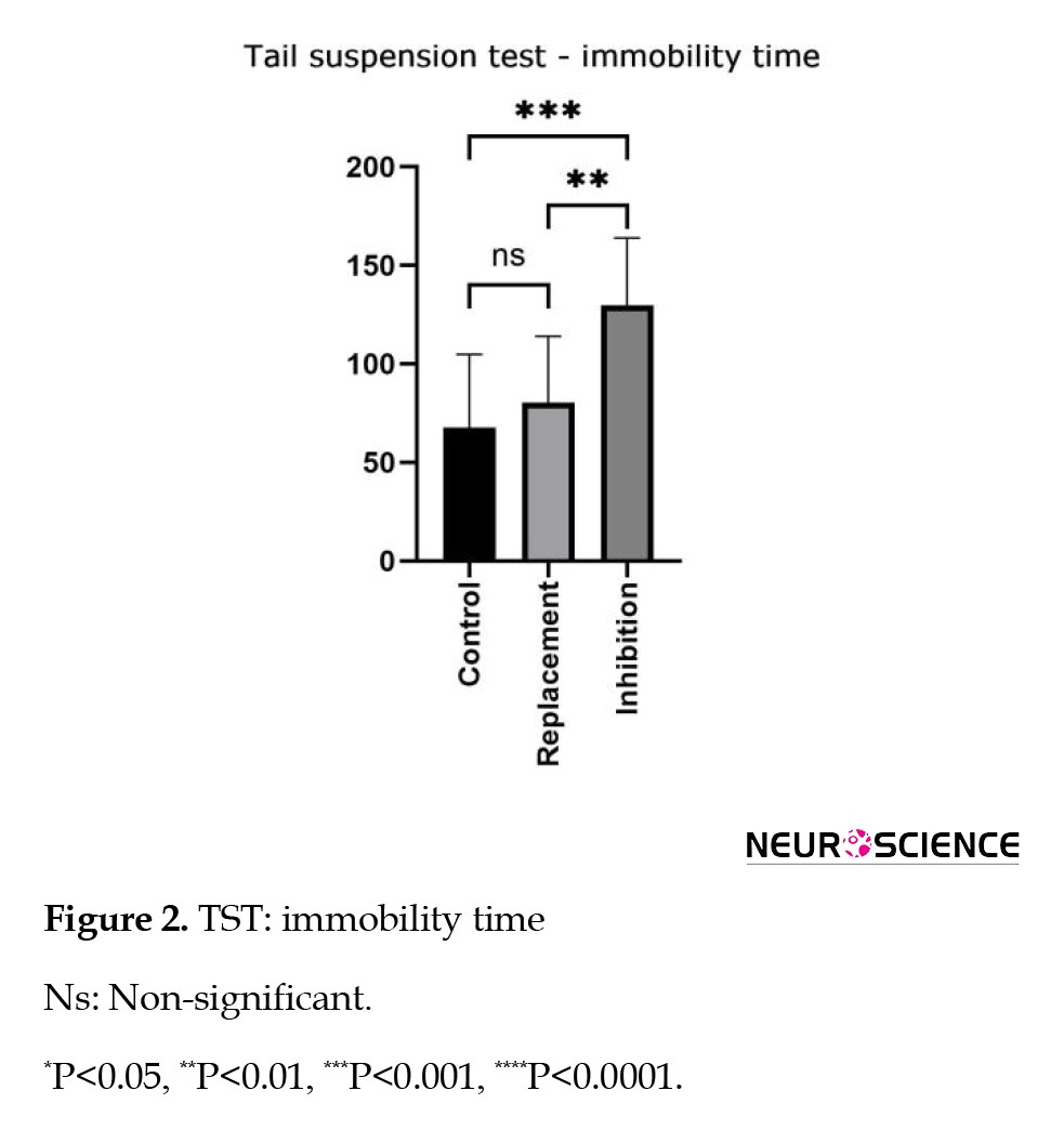
In the TST, immobility time varied significantly among groups (ANOVA, F=7.42, P=0.000398). Post-hoc analysis revealed that rats in the TSH inhibition group exhibited significantly longer immobility times compared to the blank group (P=0.0003) and the replacement group (P=0.005). No significant difference was observed between the replacement and blank groups (P=0.3255) (Figure 2).
Full-Text:
Introduction
The relationship between thyroid dysfunction and mood disorders has been extensively studied. Clinical investigations have demonstrated that patients with hyperthyroidism often exhibit symptoms, such as anxiety, irritability, and insomnia, while those with hypothyroidism are more susceptible to depression, cognitive impairment, and mental retardation (Davis et al., 2010). This suggests that the dysregulation of thyroid hormone levels not only affects peripheral metabolism but also has a profound impact on the central nervous system’s function (Ritchie & Yeap, 2015). In recent years, the increasing incidence of thyroid cancer has led to a significant rise in the number of patients undergoing total thyroidectomy. To mitigate the risk of tumor recurrence, clinical practice routinely employs thyroid-stimulating hormone (TSH) inhibition therapy, which involves the administration of high-dose exogenous levothyroxine (L-T4) to maintain serum TSH levels at an extremely low range for an extended period (Schlumberger & Leboulleux, 2021). However, growing clinical observations indicate that long-term TSH inhibition may be associated with mental health symptoms, such as anxiety and depression, which can impose a burden on patients’ quality of life (Jin et al., 2019).
The pathogenesis of emotional disorders is multifaceted, with the hippocampus playing a crucial role in regulating emotions and cognition (Jurado-Flores et al., 2022). Thyroid hormones can cross the blood-brain barrier and directly influence the activities of hippocampal neurons, thereby modulating neurotransmitter metabolism, synaptic plasticity, and neurogenesis (Ritchie & Yeap, 2015).
Existing evidence suggests that abnormal thyroid function can lead to a decrease in the level of 5-hydroxytryptamine (5-HT), a core neurotransmitter involved in emotional stability and antidepressant mechanisms. This 5-HT deficiency is considered an important pathological basis for anxiety and depression (Bauer & Whybrow, 2021). Notably, changes in 5-HT levels not only directly affect neuronal activities but also participate in the regulation of hippocampal neurogenesis by modulating the proliferation and differentiation of neural stem cells (NSCs) (Vancamp et al., 2020).
Therefore, TSH inhibition may indirectly contribute to the development of emotional disorders through the pathway of “decreased 5-HT → impaired neurogenesis”.
The Notch signaling pathway is a critical molecular network that regulates the fate determination of NSCs during neurogenesis (Ding et al., 2022). Previous studies have suggested that excessive Notch signaling is associated with decreased neuronal differentiation, increased astrocyte production, and the development of depression-like behaviors (Ran et al., 2024); for instance, in animal models of depression, Notch hyperactivation prevents the conversion of precursor cells into neurons and promotes glial lineage commitment (Ran et al., 2024). It can be hypothesized that the decrease in serotonin (5-HT) levels caused by TSH inhibition may abnormally activate the Notch pathway, disrupt the balance between neurogenesis and apoptosis, and ultimately manifest as anxiety/depression-like behaviors.
The present study employed a rat model to systematically investigate the neurobiological mechanisms underlying the association between TSH inhibition therapy and the risk of emotional disorders. Levothyroxine-induced TSH inhibition was used to establish the experimental model, and a comprehensive assessment was conducted to evaluate the effects on emotion-related behaviors, hippocampal serotonin (5-HT) levels, Notch signaling activity, and the neurogenesis/apoptosis process across behavioral, neurochemical, and molecular levels. The findings of this study aim to elucidate the potential mechanisms by which TSH inhibition may contribute to the development of emotional disorders, providing experimental evidence to inform the assessment and management of neuropsychiatric risks in clinical patients undergoing long-term TSH inhibition therapy for thyroid cancer.
Materials and Methods
Experimental animals
In this experiment, 120 SPF-grade male Wistar rats, sourced from Beijing Vital River Laboratory Animal Technology Co., Ltd. (License No. SCXK (Beijing) 2016-0006), were used. At purchase, the rats were approximately 6 weeks old, weighing 200±10 g, and measuring 12.9±1.4 cm in length. They were housed at the Laboratory Animal Center of Beijing University of Chinese Medicine under controlled conditions: 22±2 °C temperature, 50–60% humidity, and a 12-hour light/dark cycle. Standard pellet feed and water were provided ad libitum. The rats underwent a 7-day acclimation period to mitigate stress from transport and environmental changes. The study adhered to the Regulations for the Administration of Laboratory Animals and received approval from the Laboratory Animal Ethics Committee of Beijing University of Chinese Medicine.
Experimental drug
Levothyroxine (L-thyroxine, L-T4; Sigma-Aldrich, Merck, product number: T2501, 50 μg/tablet) was dissolved in sterile normal saline to achieve the desired concentration and used immediately to maintain drug efficacy.
Grouping and modeling
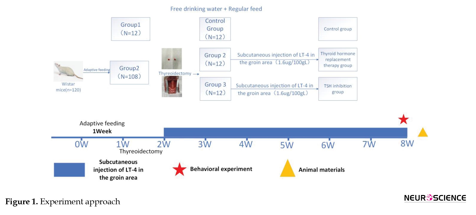
A cohort of 120 rats was randomly allocated into two groups: 12 rats served as the blank control group, while the remaining 108 underwent total thyroidectomy to establish a model, following the methodology outlined by Jin et al. (2021). Under pentobarbital sodium anesthesia (3%, 0.1 mL/100 g, intraperitoneally), bilateral thyroidectomy was performed microscopically. One week post-operation, the survival rate of the model rats was approximately 56.5%, resulting in 61 successfully modeled survivors. These 61 rats were further divided into two groups using a random number table, with 12 rats per group: the hormone replacement group and the TSH inhibition group. Both groups received daily morning drug administration for model establishment. These L-T4 doses (1.2 and 1.6 μg/100 g) were chosen based on previous studies to achieve the intended degree of TSH inhibition (Jin et al., 2021).
1. Control group: No thyroidectomy was performed. The animals were reared conventionally without any drug intervention.
2. Replacement group: Rats that underwent total thyroidectomy were subcutaneously injected with L-T4 (1.2 μg/100 g body weight) once a day for 15 consecutive days.
3. TSH inhibition group: Rats that underwent total thyroidectomy were subcutaneously injected with L-T4 (1.6 μg/100 g body weight) once a day for 15 consecutive days.
Throughout the modeling and drug administration phases, we meticulously monitored the rats’ body weight, activity levels, and overall condition to assess the impact of the modeling and interventions. The overall experimental design, animal grouping, interventions, and study timeline are summarized in Figure 1.
The relationship between thyroid dysfunction and mood disorders has been extensively studied. Clinical investigations have demonstrated that patients with hyperthyroidism often exhibit symptoms, such as anxiety, irritability, and insomnia, while those with hypothyroidism are more susceptible to depression, cognitive impairment, and mental retardation (Davis et al., 2010). This suggests that the dysregulation of thyroid hormone levels not only affects peripheral metabolism but also has a profound impact on the central nervous system’s function (Ritchie & Yeap, 2015). In recent years, the increasing incidence of thyroid cancer has led to a significant rise in the number of patients undergoing total thyroidectomy. To mitigate the risk of tumor recurrence, clinical practice routinely employs thyroid-stimulating hormone (TSH) inhibition therapy, which involves the administration of high-dose exogenous levothyroxine (L-T4) to maintain serum TSH levels at an extremely low range for an extended period (Schlumberger & Leboulleux, 2021). However, growing clinical observations indicate that long-term TSH inhibition may be associated with mental health symptoms, such as anxiety and depression, which can impose a burden on patients’ quality of life (Jin et al., 2019).
The pathogenesis of emotional disorders is multifaceted, with the hippocampus playing a crucial role in regulating emotions and cognition (Jurado-Flores et al., 2022). Thyroid hormones can cross the blood-brain barrier and directly influence the activities of hippocampal neurons, thereby modulating neurotransmitter metabolism, synaptic plasticity, and neurogenesis (Ritchie & Yeap, 2015).
Existing evidence suggests that abnormal thyroid function can lead to a decrease in the level of 5-hydroxytryptamine (5-HT), a core neurotransmitter involved in emotional stability and antidepressant mechanisms. This 5-HT deficiency is considered an important pathological basis for anxiety and depression (Bauer & Whybrow, 2021). Notably, changes in 5-HT levels not only directly affect neuronal activities but also participate in the regulation of hippocampal neurogenesis by modulating the proliferation and differentiation of neural stem cells (NSCs) (Vancamp et al., 2020).
Therefore, TSH inhibition may indirectly contribute to the development of emotional disorders through the pathway of “decreased 5-HT → impaired neurogenesis”.
The Notch signaling pathway is a critical molecular network that regulates the fate determination of NSCs during neurogenesis (Ding et al., 2022). Previous studies have suggested that excessive Notch signaling is associated with decreased neuronal differentiation, increased astrocyte production, and the development of depression-like behaviors (Ran et al., 2024); for instance, in animal models of depression, Notch hyperactivation prevents the conversion of precursor cells into neurons and promotes glial lineage commitment (Ran et al., 2024). It can be hypothesized that the decrease in serotonin (5-HT) levels caused by TSH inhibition may abnormally activate the Notch pathway, disrupt the balance between neurogenesis and apoptosis, and ultimately manifest as anxiety/depression-like behaviors.
The present study employed a rat model to systematically investigate the neurobiological mechanisms underlying the association between TSH inhibition therapy and the risk of emotional disorders. Levothyroxine-induced TSH inhibition was used to establish the experimental model, and a comprehensive assessment was conducted to evaluate the effects on emotion-related behaviors, hippocampal serotonin (5-HT) levels, Notch signaling activity, and the neurogenesis/apoptosis process across behavioral, neurochemical, and molecular levels. The findings of this study aim to elucidate the potential mechanisms by which TSH inhibition may contribute to the development of emotional disorders, providing experimental evidence to inform the assessment and management of neuropsychiatric risks in clinical patients undergoing long-term TSH inhibition therapy for thyroid cancer.
Materials and Methods
Experimental animals
In this experiment, 120 SPF-grade male Wistar rats, sourced from Beijing Vital River Laboratory Animal Technology Co., Ltd. (License No. SCXK (Beijing) 2016-0006), were used. At purchase, the rats were approximately 6 weeks old, weighing 200±10 g, and measuring 12.9±1.4 cm in length. They were housed at the Laboratory Animal Center of Beijing University of Chinese Medicine under controlled conditions: 22±2 °C temperature, 50–60% humidity, and a 12-hour light/dark cycle. Standard pellet feed and water were provided ad libitum. The rats underwent a 7-day acclimation period to mitigate stress from transport and environmental changes. The study adhered to the Regulations for the Administration of Laboratory Animals and received approval from the Laboratory Animal Ethics Committee of Beijing University of Chinese Medicine.
Experimental drug
Levothyroxine (L-thyroxine, L-T4; Sigma-Aldrich, Merck, product number: T2501, 50 μg/tablet) was dissolved in sterile normal saline to achieve the desired concentration and used immediately to maintain drug efficacy.
Grouping and modeling
A cohort of 120 rats was randomly allocated into two groups: 12 rats served as the blank control group, while the remaining 108 underwent total thyroidectomy to establish a model, following the methodology outlined by Jin et al. (2021). Under pentobarbital sodium anesthesia (3%, 0.1 mL/100 g, intraperitoneally), bilateral thyroidectomy was performed microscopically. One week post-operation, the survival rate of the model rats was approximately 56.5%, resulting in 61 successfully modeled survivors. These 61 rats were further divided into two groups using a random number table, with 12 rats per group: the hormone replacement group and the TSH inhibition group. Both groups received daily morning drug administration for model establishment. These L-T4 doses (1.2 and 1.6 μg/100 g) were chosen based on previous studies to achieve the intended degree of TSH inhibition (Jin et al., 2021).
1. Control group: No thyroidectomy was performed. The animals were reared conventionally without any drug intervention.
2. Replacement group: Rats that underwent total thyroidectomy were subcutaneously injected with L-T4 (1.2 μg/100 g body weight) once a day for 15 consecutive days.
3. TSH inhibition group: Rats that underwent total thyroidectomy were subcutaneously injected with L-T4 (1.6 μg/100 g body weight) once a day for 15 consecutive days.
Throughout the modeling and drug administration phases, we meticulously monitored the rats’ body weight, activity levels, and overall condition to assess the impact of the modeling and interventions. The overall experimental design, animal grouping, interventions, and study timeline are summarized in Figure 1.
Behavioral testing
Open field test (OFT)
The experimental setup consisted of a transparent polycarbonate cube (100×100×50 cm3), with the bottom surface divided into 9 equal-area quadrants. A 4K camera (60 Hz) was mounted at the top, and XR-SuperMaze 3.0 software was used for video recording and trajectory analysis. The experiment was conducted in a quiet environment maintained at approximately 40 lux illuminance. Each rat was placed in the center of the apparatus and allowed to move freely for 6 minutes. The recorded behavioral measures included total displacement, average speed, immobility duration (time spent completely inactive), latency to first enter the central area (an index of anxiety-related avoidance), frequency of visits to the central area, and proportion of time spent in the central area. Immediately after each trial, the apparatus was cleaned with 75% ethanol, ventilated, and dried before the next animal was tested.
Tail suspension test (TST)
The experiment took place during the dark cycle (illuminance ≤10 lux) at a room temperature of 22±1 °C and a relative humidity of 50%±5%. The rat’s tail was clamped 1±0.2 cm from the tip. Prior to the test, rats acclimated for 60 seconds before the formal timing commenced for 360 seconds. Immobility time during passive suspension was recorded and defined as the complete stillness of all four limbs, with only respiratory movements observed. In this test, a longer immobility time is interpreted as depression-like behavior.
Specimen preparation
Serum preparation
Following a 12-hour fast, the rats were weighed and anesthetized via an intraperitoneal injection of 3% pentobarbital sodium (0.1 mL/100 g). Once anesthesia was confirmed, the abdominal aorta was exposed, and approximately 5 mL of blood was drawn using a vacuum blood collection tube, completing the procedure within 5 minutes. The blood samples stood for 30 minutes before being centrifuged at 3000 r/min for 10 minutes to separate the serum, which was then stored at –80 °C for future analysis.
Brain tissue collection
Following blood collection, the rats were sacrificed by decapitation, and the entire brain was extracted within 120 seconds (to minimize post-mortem tissue changes). The brains were placed on a dissection table cooled to 4 °C. Under these low-temperature conditions, the surface was rinsed with sterile saline to eliminate residual blood. The hippocampal region was meticulously dissected under a binocular microscope, divided into aliquots in pre-frozen 2 mL cryotubes, labeled, and promptly stored at –80 °C.
Enzyme-linked immunosorbent assay (ELISA)
Serum thyroid function indices (TSH, FT3, and FT4) and hippocampal 5-HT levels were quantified using commercial ELISA kits, following the manufacturers’ protocols. Optical density values at 450 nm were measured with a Bio-Rad Model 680 microplate reader, and a standard curve was constructed for quantitative analysis.
Double-label immunofluorescence
Hippocampal tissues were fixed in 4% paraformaldehyde, dehydrated with 30% sucrose, and sectioned into 10 μm paraffin slices. Following antigen retrieval and blocking, primary antibodies were applied and incubated overnight at 4 °C. The next day, tissues were washed with PBS, then incubated with fluorescently labeled secondary antibodies for 1 hour at room temperature in the dark. Nuclei were stained with DAPI, visualized under a fluorescence microscope, and the number and distribution of positive cells were analyzed using ImageJ software.
Quantitative real-time PCR (qRT-PCR)
Total RNA from the hippocampus was extracted via the Trizol method, with purity and concentration assessed by Nanodrop. cDNA synthesis was conducted through reverse transcription, followed by qPCR using SYBR Green Master Mix in a 20 μL reaction system over 40 cycles. The target genes were pivotal to the Notch signaling pathway, with GAPDH serving as the internal reference. Relative expression levels were calculated using the 2-ΔΔCt method.
Western blot
Hippocampal tissues were homogenized and lysed using RIPA buffer with PMSF, followed by centrifugation at 12,000 g for 10 minutes to collect the supernatant. Protein concentration was assessed via the BCA method. Proteins (30 μg) were separated by SDS-PAGE, transferred to a membrane, and blocked with 5% non-fat milk for 1 hour. Primary antibodies were applied and incubated overnight at 4 °C. After washing with TBST, HRP-labeled secondary antibodies were added and incubated for 1 hour at room temperature. Bands were visualized using ECL and captured with a ChemiDoc MP imaging system. Quantitative analysis was conducted with ImageJ, normalizing relative expression levels to β-actin.
Statistical analysis
Data are presented as Mean±SD. Normality and homogeneity of variances were assessed prior to analysis. One-way analysis of variance (ANOVA) was used for comparisons among multiple groups, followed by the least significant difference (LSD) method for pairwise comparisons. Non-parametric tests (e.g. Kruskal–Wallis) were employed if the data did not conform to the normal distribution, with Dunn’s test for post-hoc multiple comparisons. Statistical analyses were performed using SPSS software, version 26.0, and figures were generated with GraphPad Prism software, version 10. The threshold for statistical significance was set at P<0.05.
Results
Behavioral performance
In the TST, immobility time varied significantly among groups (ANOVA, F=7.42, P=0.000398). Post-hoc analysis revealed that rats in the TSH inhibition group exhibited significantly longer immobility times compared to the blank group (P=0.0003) and the replacement group (P=0.005). No significant difference was observed between the replacement and blank groups (P=0.3255) (Figure 2).
This prolonged immobility indicates a heightened depression-like state in the TSH-suppressed rats.
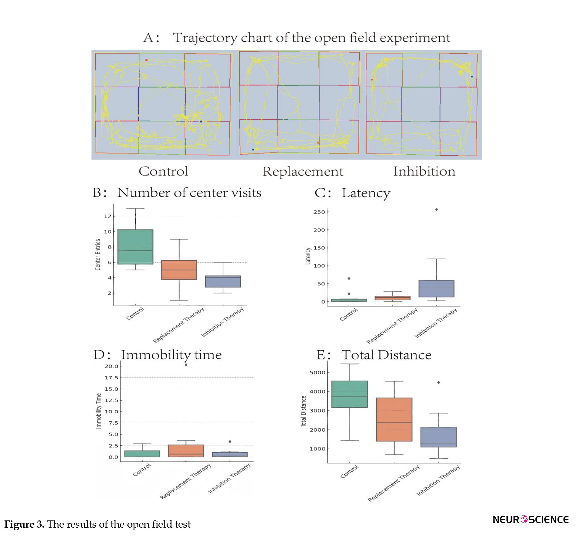
The OFT revealed distinct behavioral patterns in the inhibition group compared to controls (Figure 3).
The OFT revealed distinct behavioral patterns in the inhibition group compared to controls (Figure 3).
Rats in the inhibition group exhibited a significant reduction in the number of entries into the central arena (P<0.0001, Figure 3B). Additionally, these animals displayed a marked prolongation in the latency to enter the central area (P<0.0001, Figure 3C) and the time to first enter the central zone (P=0.0002, Figure 3D). Furthermore, the total distance traveled by the inhibition group was significantly decreased (P<0.0001, Figure 3E). Together, these OFT changes are indicative of increased anxiety-like behavior under TSH inhibition.
Changes in thyroid axis function
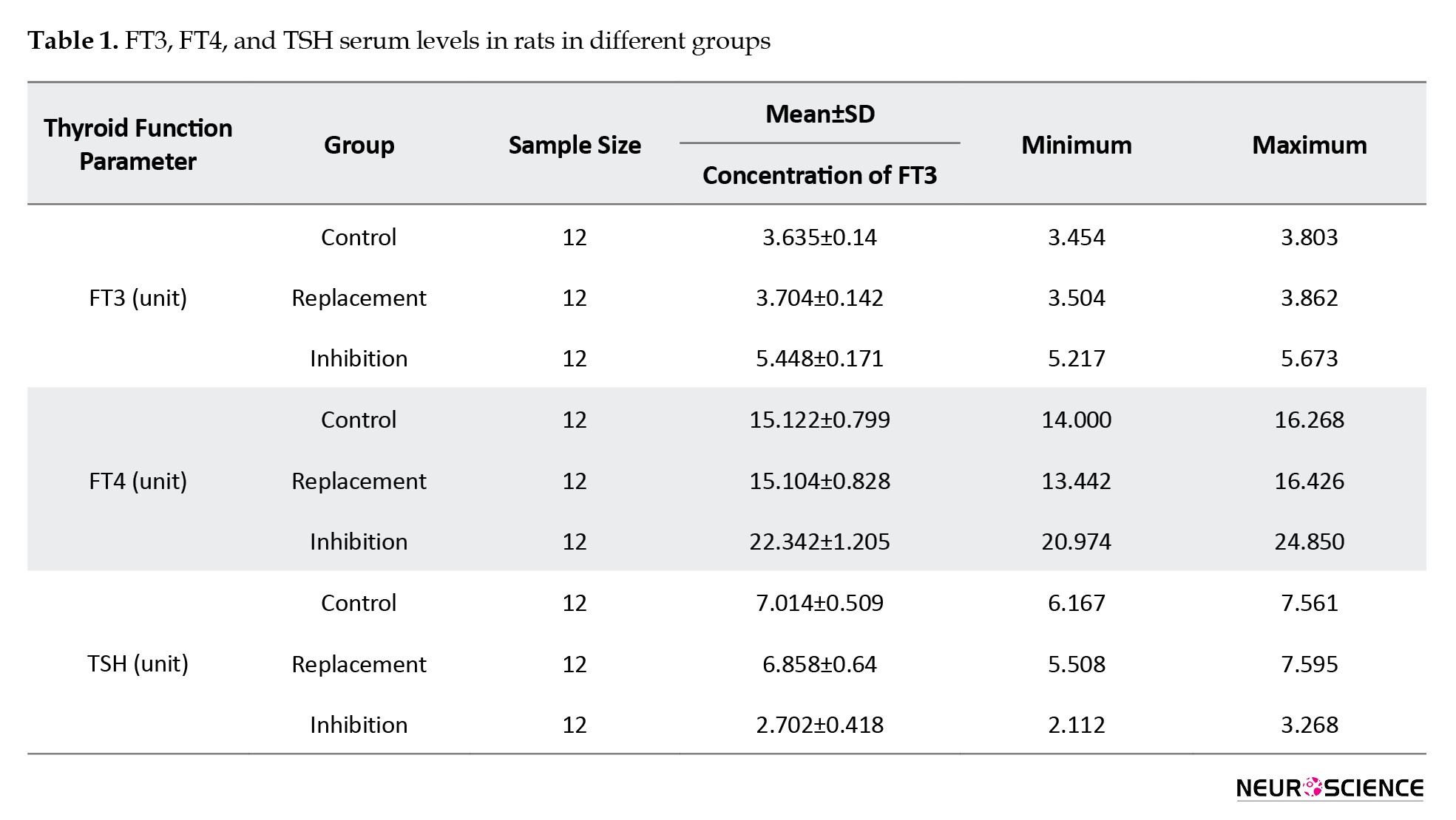
ELISA assay results demonstrated that TSH inhibition markedly influenced thyroid hormone levels. In the inhibition group, FT3 and FT4 levels were significantly elevated compared to the blank group (P<0.0001), whereas no significant difference was observed between the replacement and blank groups. TSH levels significantly decreased in the inhibition group (P<0.0001), with no significant difference between the replacement and blank groups (Table 1).
HT levels in the hippocampus
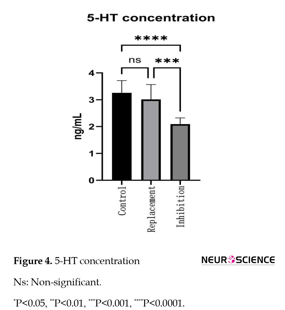
The ELISA assay revealed a significant reduction in hippocampal 5-HT content in the inhibition group compared to the blank group (1.916±0.37 vs 3.415±0.42, P<0.0001). Conversely, the replacement group exhibited no statistically significant difference from the blank group (P=0.054) (Figure 4).
Changes in thyroid axis function
ELISA assay results demonstrated that TSH inhibition markedly influenced thyroid hormone levels. In the inhibition group, FT3 and FT4 levels were significantly elevated compared to the blank group (P<0.0001), whereas no significant difference was observed between the replacement and blank groups. TSH levels significantly decreased in the inhibition group (P<0.0001), with no significant difference between the replacement and blank groups (Table 1).
HT levels in the hippocampus
The ELISA assay revealed a significant reduction in hippocampal 5-HT content in the inhibition group compared to the blank group (1.916±0.37 vs 3.415±0.42, P<0.0001). Conversely, the replacement group exhibited no statistically significant difference from the blank group (P=0.054) (Figure 4).
This pronounced decrease in hippocampal 5-HT in TSH-suppressed rats is consistent with a serotonin-mediated mechanism for the observed mood disturbances.
Changes in hippocampal neurogenesis
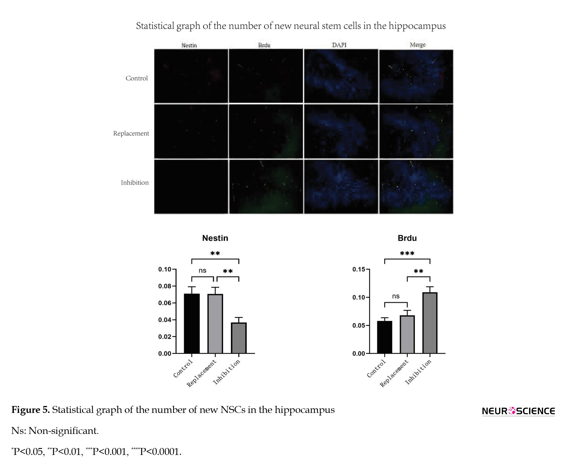
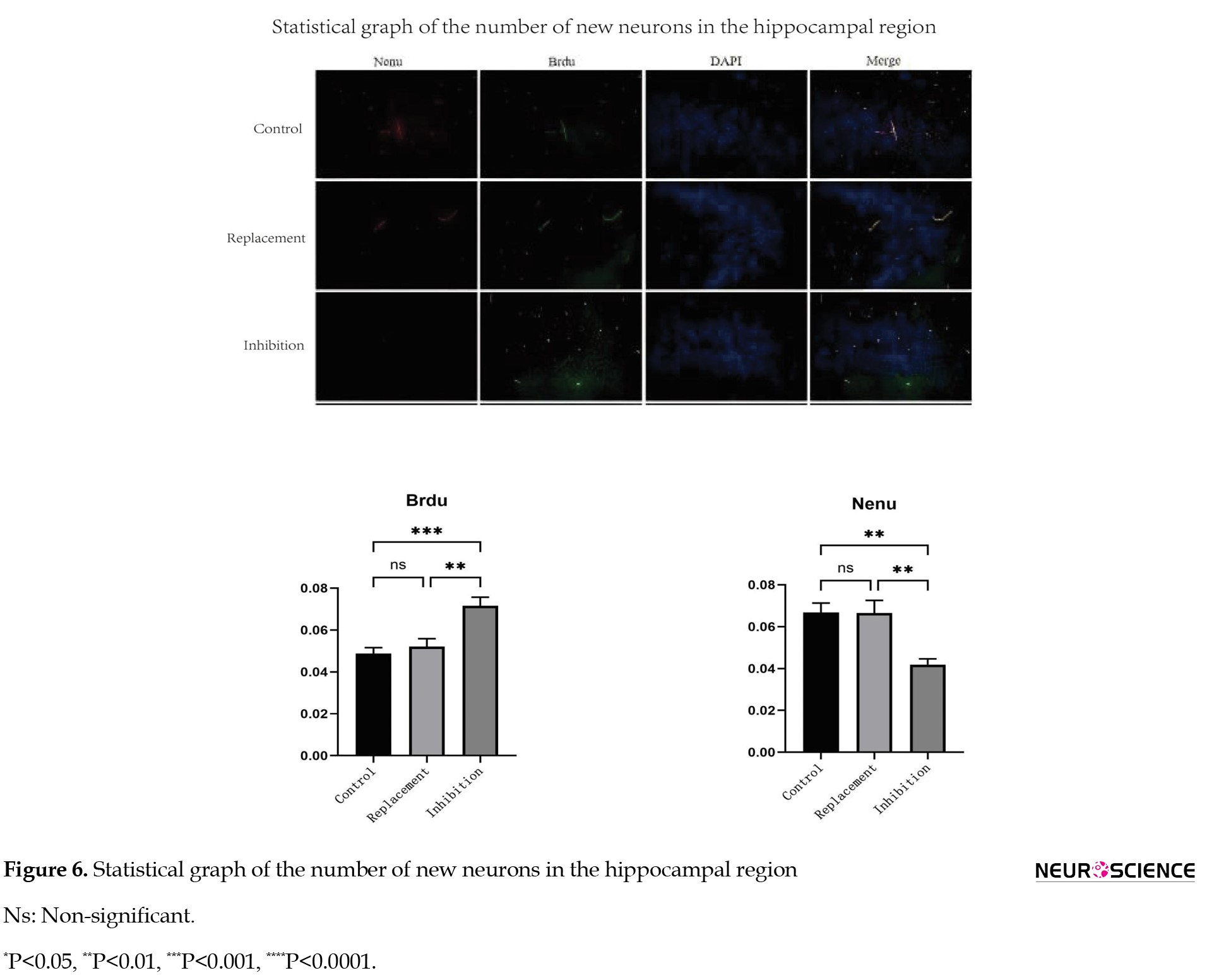
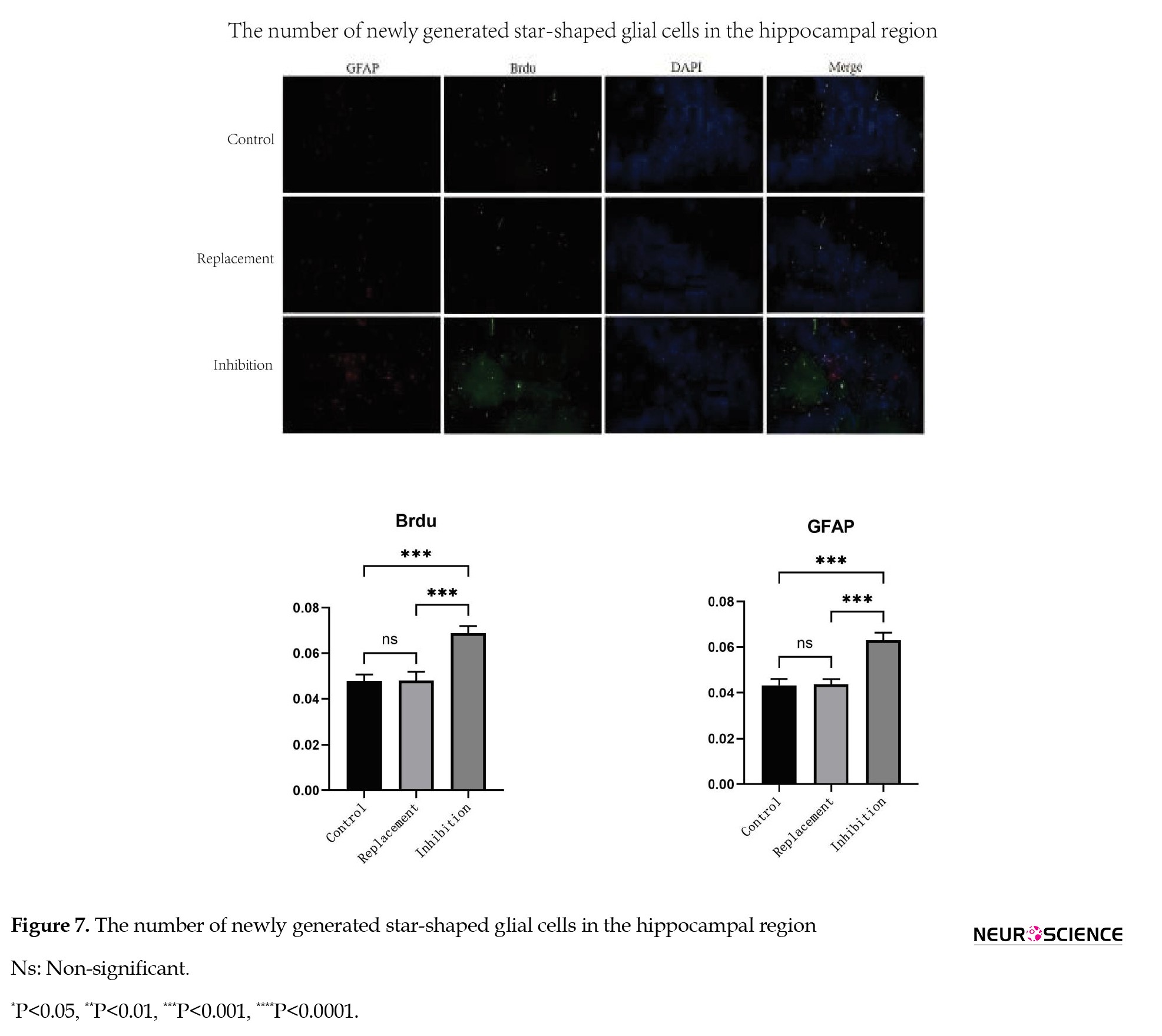
Double immunofluorescence staining revealed that, compared to the blank and replacement groups, the inhibition group exhibited significantly elevated levels of BrdU/Nestin (NSCs; P=0.0013 and P=0.0014, respectively; Figure 5), significantly reduced levels of BrdU/NeuN (newborn neurons; P=0.0006; Figure 6), and significantly increased levels of BrdU/GFAP (newborn astrocytes; P=0.0008 and P=0.0009, respectively; Figure 7).
Changes in hippocampal neurogenesis
Double immunofluorescence staining revealed that, compared to the blank and replacement groups, the inhibition group exhibited significantly elevated levels of BrdU/Nestin (NSCs; P=0.0013 and P=0.0014, respectively; Figure 5), significantly reduced levels of BrdU/NeuN (newborn neurons; P=0.0006; Figure 6), and significantly increased levels of BrdU/GFAP (newborn astrocytes; P=0.0008 and P=0.0009, respectively; Figure 7).
Overall, these findings indicate that TSH inhibition increases neural progenitor proliferation but impairs neuronal differentiation while promoting astrocytic differentiation.
Activity of the Notch signaling pathway
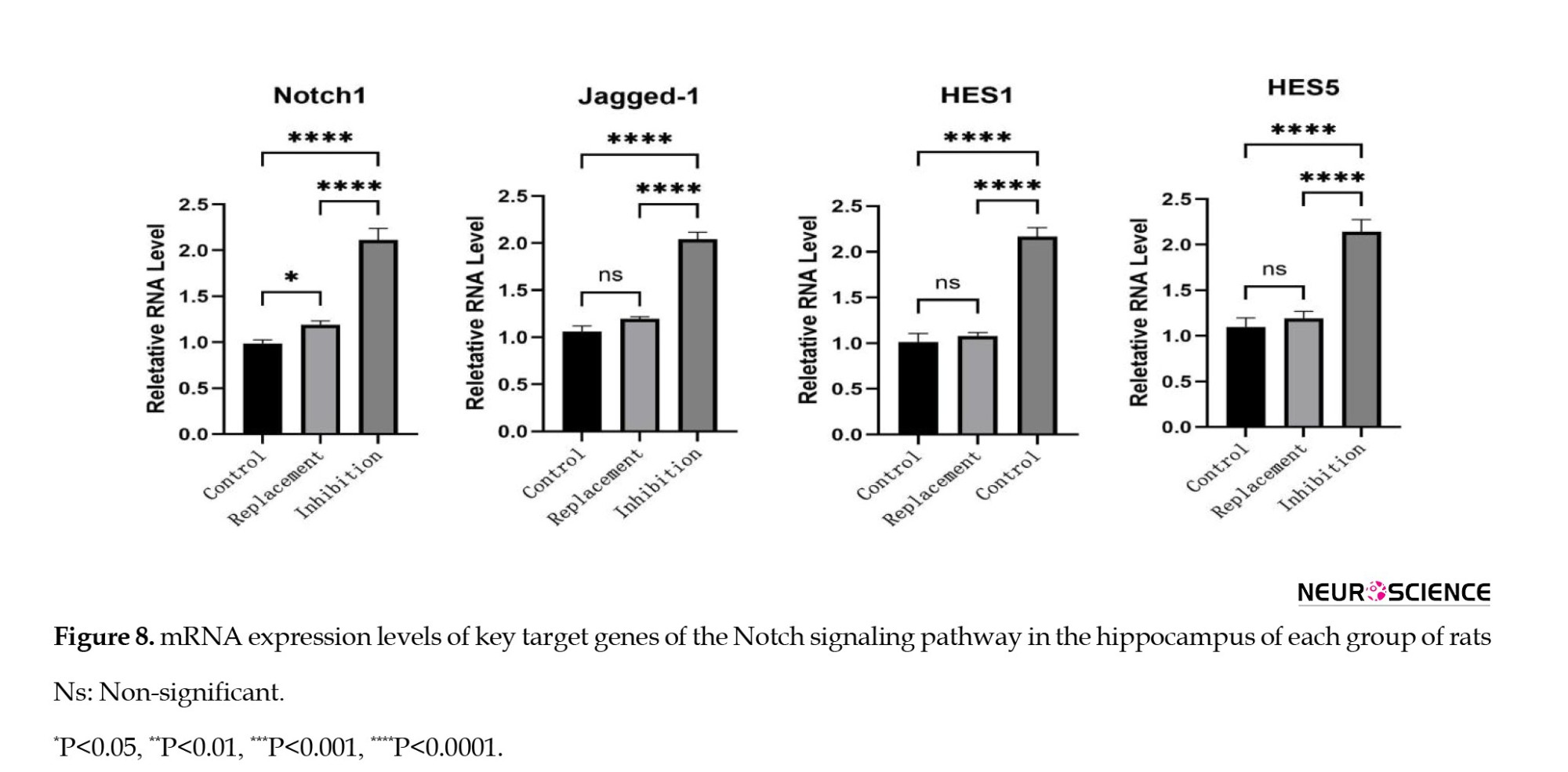
Quantitative polymerase chain reaction (qPCR) analysis revealed significant upregulation of Notch1, Hes1, Hes5, and Jagged-1 mRNA expression in the inhibition group compared to controls (all P<0.05, Figure 8).
Activity of the Notch signaling pathway
Quantitative polymerase chain reaction (qPCR) analysis revealed significant upregulation of Notch1, Hes1, Hes5, and Jagged-1 mRNA expression in the inhibition group compared to controls (all P<0.05, Figure 8).
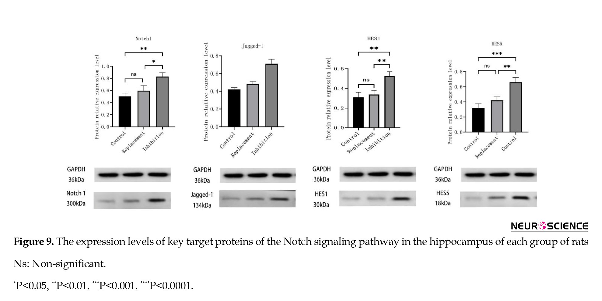
Western blotting further corroborated this trend, with Notch1, HES1, HES5, and Jagged-1 protein levels demonstrating a statistically significant increase in the inhibition group relative to the blank and replacement groups (Notch1: F=14.69, P=0.0012; HES5: F=23.04, P=0.0002; HES1: F=14.59, P=0.0013; Jagged-1: F=26.60, P=0.0002, Figure 9).
The activation of the Notch pathway in TSH-suppressed rats provides a molecular link to the observed alterations in neurogenesis.
Changes in apoptosis-related molecules
At the mRNA level, the Bax/Bcl-2 ratio was highest in the inhibition group (6.511±0.804) and was significantly greater than both the control (1.257±0.243) and replacement (2.106±0.088) groups (P<0.01 for both comparisons, Table 2).
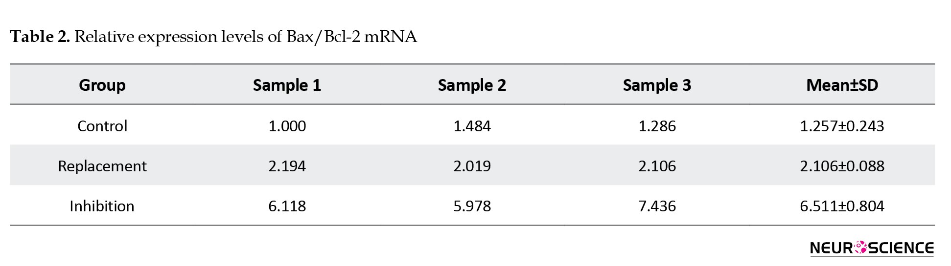
Also, mRNA levels in the replacement group were significantly higher than in the control group (P<0.01).
At the protein level, the inhibition group likewise exceeded both the control and replacement groups (0.867±0.015 vs 0.667±0.015 and 0.677±0.015, respectively; P<0.01 for both, Table 3).
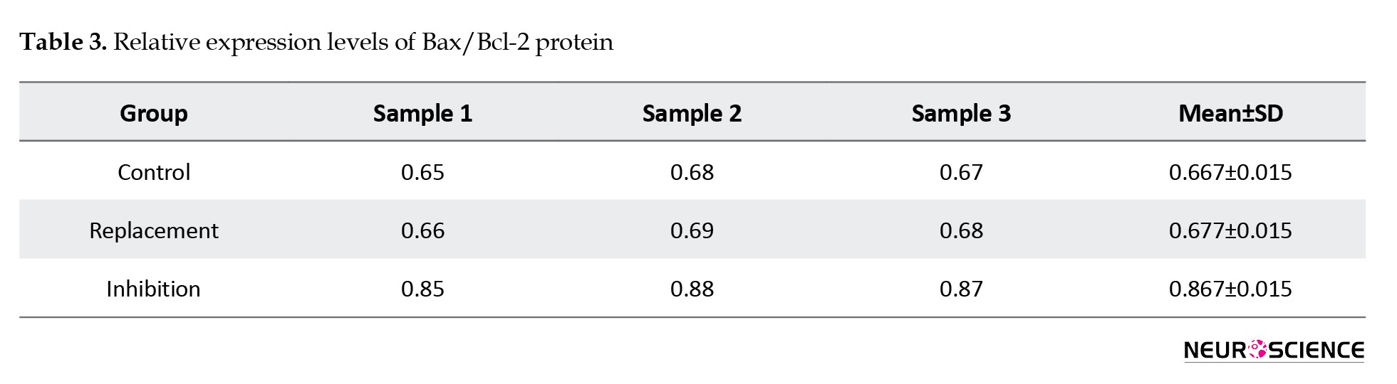
No significant difference was observed between the control and replacement groups (P>0.05). Relative to controls, the protein Bax/Bcl-2 ratio increased by ~30% in the inhibition group, indicating enhanced apoptotic signaling in the hippocampus under TSH inhibition.
Discussion
This study systematically demonstrated that prolonged TSH inhibition elicits substantial metabolic and endocrine alterations, as well as anxiety- and depression-like behaviors, by modulating hippocampal neurotransmitters, neurogenesis, and apoptosis in a rat model of TSH inhibition. These findings offer an experimental foundation for understanding the mental health symptoms experienced by clinical thyroid cancer patients following TSH inhibition therapy, and suggest potential underlying molecular mechanisms.
The present study showed that rats subjected to TSH inhibition exhibited distinct behavioral phenotypes characteristic of depression and anxiety. Specifically, these animals displayed prolonged immobility in the TST and reduced exploration with increased latency in the OFT. This finding aligns with clinical observations in thyroid cancer patients undergoing long-term TSH inhibition therapy (often maintained over months or years), who frequently report symptoms, such as anxiety, depression, and emotional instability (Botella-Carretero, 2003; Roerink et al., 2013; Jung & Visovatti, 2017). Importantly, the control group receiving hormone replacement therapy maintained euthyroid status and did not exhibit significant emotional disturbances. These results suggest that the emotional abnormalities observed are not simply a consequence of thyroid hormone deficiency, but are directly linked to the persistent low levels of TSH and the artificial disruption of the thyroid axis regulation.
The present study provides further mechanistic insights into the observed behavioral alterations associated with TSH inhibition. The findings indicated that TSH inhibition was accompanied by a significant decrease in 5-HT levels within the hippocampus. This is noteworthy, as extensive evidence has demonstrated that thyroid hormones can regulate the metabolism and transport of monoamine neurotransmitters, including 5-HT, in the brain, and the impact on the 5-HT system is particularly crucial (Kotkowska & Strzelecki, 2022). Serotonin is not only the classic neurotransmitter basis for emotional disorders, but its deficiency has also been linked to inhibited neurogenesis and reduced neural plasticity (Kamyshna et al., 2024). In the current study, the decrease in 5-HT levels coincided with the observed behavioral abnormalities, suggesting a pivotal upstream role of 5-HT dysregulation in the emotional disturbances caused by TSH inhibition. Importantly, clinical studies have reported similar findings, with hyperthyroid patients exhibiting 5-HT metabolic disorders (Mai et al., 2023) and hypothyroid patients showing decreased 5-HT synthesis and release (Osnaya-Brizuela et al., 2024), both of which are strongly correlated with depressive and anxious symptoms. Collectively, these results indicate that TSH inhibition may lead to a hormonal imbalance by over-activating the thyroid axis, which in turn alters the homeostasis of the 5-HT system, ultimately resulting in emotional abnormalities.
TSH inhibition resulted in a significant imbalance in hippocampal neurogenesis, characterized by enhanced proliferation, restricted neuronal differentiation, and increased glial differentiation. Hippocampal neurogenesis is a core pathological mechanism implicated in emotional disorders. Previous animal studies have demonstrated that both chronic stress and depression models are accompanied by reduced hippocampal neuron generation and enhanced glial reactivity (Ahmed et al., 2012). The abnormal neurogenesis pattern reported here is highly consistent with the decline in 5-HT signaling, as 5-HT can directly regulate the fate determination of NSCs. Diminished 5-HT not only weakens neuronal differentiation signals but may also lead to functional impairment of neural circuits by triggering a deviation toward the glial lineage (Baghcheghi et al., 2018). The present findings demonstrate that the Notch signaling pathway was significantly upregulated in the TSH inhibition group, as evidenced by the increased expression of Notch1, Hes1, Hes5, and Jagged-1. The Notch signaling cascade is a crucial regulator of NSC maintenance and neuronal differentiation inhibition. Its overactivation prevents the conversion of precursor cells into the neuronal lineage and promotes their glial differentiation (Xu et al., 2023). Previous research has established the abnormal hyperactivation of Notch signaling in individuals with depression and animal models of chronic stress, which is accompanied by restricted hippocampal neurogenesis (Maglione et al., 2022). The current results not only corroborate this phenomenon but also suggest that TSH inhibition, through the decrease of upstream 5-HT, may interact with the imbalance of Notch signaling, ultimately leading to impaired neurogenesis.
This study identified increased apoptosis as a significant finding alongside impaired neurogenesis. Rats subjected to TSH inhibition displayed a higher Bax/Bcl-2 ratio, indicating a pro-apoptotic state. The Bax/Bcl-2 ratio was approximately 30% higher in TSH-suppressed rats than in controls, confirming this heightened apoptosis. Previous research links abnormal thyroid function to oxidative stress and mitochondrial damage, accelerating neuronal apoptosis (Liao et al., 2021). The combined impact of heightened neuronal apoptosis and impaired neurogenesis compromises hippocampal plasticity and functionality, contributing to emotional disorders (Schroer et al., 2023). Mechanistically, reduced 5-HT levels not only diminish neuronal survival but may also disrupt Notch signaling and increase apoptosis by impairing BDNF signaling (Ru et al., 2024). Consequently, emotional disorders may arise from intertwined pathological processes: hormonal imbalance reduces 5-HT, impairing neurogenesis via Notch signaling and apoptotic pathways, ultimately leading to anxiety- and depression-like outcomes in behavior. It should be noted that other mechanisms not extensively examined here, such as neuroinflammation and impaired neurotrophic support (e.g. reduced BDNF/TrkB signaling), might also contribute to these neuropsychiatric effects of TSH inhibition, and deserve exploration in future studies.
This study elucidates a comprehensive mechanism: TSH inhibition disrupts thyroid hormone levels, leading to reduced hippocampal 5-HT, aberrant Notch pathway activation, and restricted neuronal differentiation. This results in increased glial differentiation, heightened apoptosis signals, and reduced neural network plasticity, culminating in depressive and anxiety-like behaviors. Aligning with previous clinical findings, this mechanism offers a robust experimental foundation for understanding mood disorders associated with TSH inhibition therapy, highlighting potential neuropsychiatric risks of excessive inhibition.
This study has several limitations that warrant consideration. First, the extent and temporal dynamics of TSH inhibition may not fully recapitulate clinical scenarios, underscoring the need for cautious extrapolation of the results from animal models to human patients. Second, the investigation primarily focused on the Notch signaling pathway, while other potentially relevant mechanisms, such as neuroinflammation, oxidative stress, the BDNF/TrkB pathway, and alterations in the hypothalamic-pituitary-adrenal axis, were not comprehensively examined. Finally, the current findings are correlative in nature, and future pharmacological or genetic manipulations will be necessary to establish the causal relationship between reduced serotonin levels and dysregulation of the Notch pathway. Conducting a longitudinal study in clinical populations that integrates serological, neuroimaging, and cognitive assessments could help clarify the predictive value of TSH inhibition therapy for the risk of emotional disorders, thereby informing personalized treatment approaches.
In summary, this study elucidates the mechanisms by which TSH inhibition may lead to emotional disorders, examining behavioral science, neurotransmitters, neurogenesis, and molecular signaling pathways. The findings indicated that prolonged TSH inhibition poses risks, necessitating a balance between tumor control and mental health. Further clarification of the 5-HT–Notch–neurogenesis pathway and exploration of drug interventions could inform comprehensive management strategies for thyroid cancer patients.
Conclusion
TSH inhibition is associated with anxiety/depression-like behaviors, accompanied by a decrease in hippocampal 5-HT levels, an upregulation of the Notch pathway, and an imbalance in neurogenesis–apoptosis. It suggests that excessive inhibition of the thyroid axis may pose neuropsychiatric risks, and a balance needs to be struck between tumor recurrence inhibition and mental health. These results highlight the importance of routine psychological monitoring and support for patients undergoing long-term TSH inhibition therapy.
Ethical Considerations
Compliance with ethical guidelines
This study adhered to the Regulations for the Administration of Laboratory Animals and received approval from the Laboratory Animal Ethics Committee of Beijing University of Chinese Medicine, Beijing, China (Code: BUCM-2024060102-2228).
Funding
This work was supported by the National Natural Science Foundation of China, Beijing, China (Grant No.: 81973855); the Clinical Research Business Expenses of Central High-level Hospitals at the provincial-ministerial level (Research No.: 2022-NHLHCRF-LX-02-0115).
Authors' contributions
Conceptualization, project administration, and writing: Yikun Zhao and Honglin Jiang; Validation: Huiyuan Shang; Investigation and final approval: All authors.
Conflict of interest
The authors declared no conflict of interest.
References
Ahmed, O. M., Ahmed, R. G., El-Gareib, A. W., El-Bakry, A. M., & Abd El-Tawab, S. M. (2012). Effects of experimentally induced maternal hypothyroidism and hyperthyroidism on the development of rat offspring: II-the developmental pattern of neurons in relation to oxidative stress and antioxidant defense system. International Journal of Developmental Neuroscience: The Official Journal of the International Society for Developmental Neuroscience, 30(6), 517–537. [DOI:10.1016/j.ijdevneu.2012.04.005] [PMID]
Baghcheghi, Y., Beheshti, F., Shafei, M. N., Salmani, H., Sadeghnia, H. R., & Soukhtanloo, M., et al. (2018). The effects of vitamin E on brain derived neurotrophic factor, tissues oxidative damage and learning and memory of juvenile hypothyroid rats. Metabolic Brain Disease, 33(3), 713–724. [DOI:10.1007/s11011-017-0176-0] [PMID]
Bauer, M., & Whybrow, P. C. (2021). Role of thyroid hormone therapy in depressive disorders. Journal of Endocrinological Investigation, 44(11), 2341–2347. [DOI:10.1007/s40618-021-01600-w] [PMID]
Botella-Carretero, J. I., Galán, J. M., Caballero, C., Sancho, J., & Escobar-Morreale, H. F. (2003). Quality of life and psychometric functionality in patients with differentiated thyroid carcinoma. Endocrine-Related Cancer, 10(4), 601–610. [DOI:10.1677/erc.0.0100601] [PMID]
Davis, P. J., Zhou, M., Davis, F. B., Lansing, L., Mousa, S. A., & Lin, H. Y. (2010). Mini-review: Cell surface receptor for thyroid hormone and nongenomic regulation of ion fluxes in excitable cells. Physiology & Behavior, 99(2), 237–239. [DOI:10.1016/j.physbeh.2009.02.015] [PMID]
Ding, M. R., Qu, Y. J., Hu, B., & An, H. M. (2022). Signal pathways in the treatment of Alzheimer's disease with traditional Chinese medicine. Biomedicine & Pharmacotherapy = Biomedecine & Pharmacotherapie, 152, 113208. [DOI:10.1016/j.biopha.2022.113208] [PMID]
Jin, S., & Sugitani, I. (2021). Development of a rat model for evaluating thyroid-stimulating hormone suppression after total thyroidectomy. Journal of Nippon Medical School = Nippon Ika Daigaku zasshi, 88(4), 311–318. [DOI:10.1272/jnms.JNMS.2021_88-409] [PMID]
Jin, S., Yang, Y. T., Bao, W., Bai, Y., Ai, J. W., & Liu, Y., et al. (2019). Naming difficulties after thyroid stimulating hormone suppression therapy in patients with differentiated thyroid carcinoma: A prospective cohort study. Endocrine, 65(2), 327–337. [DOI:10.1007/s12020-019-01943-8] [PMID]
Jung, M. S., & Visovatti, M. (2017). Post-treatment cognitive dysfunction in women treated with thyroidectomy for papillary thyroid carcinoma. Supportive Care in Cancer: Official Journal of the Multinational Association of Supportive Care in Cancer, 25(3), 915–923. [DOI:10.1007/s00520-016-3481-y] [PMID]
Jurado-Flores, M., Warda, F., & Mooradian, A. (2022). pathophysiology and clinical features of neuropsychiatric manifestations of thyroid disease. Journal of the Endocrine Society, 6(2), bvab194. [DOI:10.1210/jendso/bvab194] [PMID]
Kamyshna, I., Pavlovych, L., Pankiv, V., Khodorovska, A., Bilous, O., & Kamyshnyi, O. (2024). The molecular fundamentals of neurorehabilitation and their modulation by thyroid hormones. International Journal of Endocrinology (Ukraine), 20(2), 126–132. [DOI:10.22141/2224-0721.20.2.2024.1374]
Kotkowska, Z., & Strzelecki, D. (2022). Depression and autoimmune hypothyroidism-their relationship and the effects of treating psychiatric and thyroid disorders on changes in clinical and biochemical parameters including BDNF and other cytokines-a systematic review. Pharmaceuticals (Basel, Switzerland), 15(4), 391. [DOI:10.3390/ph15040391] [PMID]
Liao, J., Zeng, G., Fang, W., Huang, W., Dai, X., & Ye, Q., et al. (2021). Increased Notch2/NF-κB signaling may mediate the depression susceptibility: Evidence from chronic social defeat stress mice and WKY rats. Physiology & Behavior, 228, 113197. [DOI:10.1016/j.physbeh.2020.113197] [PMID]
Maglione, A. V., do Nascimento, B. P. P., Ribeiro, M. O., de Souza, T. J. L., da Silva, R. E. C., & Sato, M. A., et al. (2022). Triiodothyronine treatment reverses depression-like behavior in a triple-transgenic animal model of Alzheimer's disease. Metabolic Brain Disease, 37(8), 2735–2750. [DOI:10.1007/s11011-022-01055-9] [PMID]
Mai, A., Essam, R. M., Ahmed, K. A., & Abdelmonem, M. (2023). Implication of Wnt/GSK-3β/β-Catenin Signaling in the pathogenesis of mood disturbances associated with hyperthyroidism in rats: Potential therapeutic effect of Naringin. ACS Chemical Neuroscience, 14(11), 2035–2048. [PMID]
Osnaya-Brizuela, N., Valenzuela-Peraza, A., Santamaría-Del Ángel, D., García-Martínez, Y., Pacheco-Rosado, J., & Pérez-Sánchez, G., et al. (2024). Is the acquired hypothyroidism a risk factor for developing psychiatric disorders? Frontiers in Psychiatry, 15, 1429255. [DOI:10.3389/fpsyt.2024.1429255] [PMID]
Ran, L. C., Shang, H., Yuan, Y., Chen, L. R., Yang, T., & Li, L., et al. (2024). [Mechanism of electroacupuncture on hippocampal neuron regeneration in depression rats based on Wnt/β-catenin and Notch signaling pathway]. Zhen ci yan jiu = Acupuncture Research, 49(12), 1282–1288. [PMID]
Ritchie, M., & Yeap, B. B. (2015). Thyroid hormone: Influences on mood and cognition in adults. Maturitas, 81(2), 266–275. [DOI:10.1016/j.maturitas.2015.03.016] [PMID]
Roerink, S. H., de Ridder, M., Prins, J., Huijbers, A., de Wilt, H. J., & Marres, H., et al. (2013). High level of distress in long-term survivors of thyroid carcinoma: Results of rapid screening using the distress thermometer. Acta Oncologica (Stockholm, Sweden), 52(1), 128–137. [DOI:10.3109/0284186X.2012.723822] [PMID]
Ru, D., Zhang, Z., Liu, M., Fan, X., Wang, Y., & Yan, Y., et al. (2024). Downregulation of notch signaling-stimulated genes in neurovascular unit alterations induced by chronic cerebral hypoperfusion. Immunity, Inflammation and Disease, 12(11), e70082. [DOI:10.1002/iid3.70082] [PMID]
Schlumberger, M., & Leboulleux, S. (2021). Current practice in patients with differentiated thyroid cancer. Nature reviews. Endocrinology, 17(3), 176–188. [DOI:10.1038/s41574-020-00448-z] [PMID]
Schroer, J., Warm, D., De Rosa, F., Luhmann, H. J., & Sinning, A. (2023). Activity-dependent regulation of the BAX/BCL-2 pathway protects cortical neurons from apoptotic death during early development. Cellular and Molecular Life Sciences: CMLS, 80(6), 175. [DOI:10.1007/s00018-023-04824-6] [PMID]
Vancamp, P., Butruille, L., Demeneix, B. A., & Remaud, S. (2020). Thyroid hormone and neural stem cells: repair potential following brain and spinal cord injury. Frontiers in Neuroscience, 14, 875. [DOI:10.3389/fnins.2020.00875] [PMID]
Xu, Y., Yang, L., Teng, Y., Li, J., & Li, N. (2023). Exploring the underlying molecular mechanism of tri(1,3-dichloropropyl) phosphate-induced neurodevelopmental toxicity via thyroid hormone disruption in zebrafish by multi-omics analysis. Aquatic Toxicology (Amsterdam, Netherlands), 258, 106510. [DOI:10.1016/j.aquatox.2023.106510] [PMID]
Changes in apoptosis-related molecules
At the mRNA level, the Bax/Bcl-2 ratio was highest in the inhibition group (6.511±0.804) and was significantly greater than both the control (1.257±0.243) and replacement (2.106±0.088) groups (P<0.01 for both comparisons, Table 2).
Also, mRNA levels in the replacement group were significantly higher than in the control group (P<0.01).
At the protein level, the inhibition group likewise exceeded both the control and replacement groups (0.867±0.015 vs 0.667±0.015 and 0.677±0.015, respectively; P<0.01 for both, Table 3).
No significant difference was observed between the control and replacement groups (P>0.05). Relative to controls, the protein Bax/Bcl-2 ratio increased by ~30% in the inhibition group, indicating enhanced apoptotic signaling in the hippocampus under TSH inhibition.
Discussion
This study systematically demonstrated that prolonged TSH inhibition elicits substantial metabolic and endocrine alterations, as well as anxiety- and depression-like behaviors, by modulating hippocampal neurotransmitters, neurogenesis, and apoptosis in a rat model of TSH inhibition. These findings offer an experimental foundation for understanding the mental health symptoms experienced by clinical thyroid cancer patients following TSH inhibition therapy, and suggest potential underlying molecular mechanisms.
The present study showed that rats subjected to TSH inhibition exhibited distinct behavioral phenotypes characteristic of depression and anxiety. Specifically, these animals displayed prolonged immobility in the TST and reduced exploration with increased latency in the OFT. This finding aligns with clinical observations in thyroid cancer patients undergoing long-term TSH inhibition therapy (often maintained over months or years), who frequently report symptoms, such as anxiety, depression, and emotional instability (Botella-Carretero, 2003; Roerink et al., 2013; Jung & Visovatti, 2017). Importantly, the control group receiving hormone replacement therapy maintained euthyroid status and did not exhibit significant emotional disturbances. These results suggest that the emotional abnormalities observed are not simply a consequence of thyroid hormone deficiency, but are directly linked to the persistent low levels of TSH and the artificial disruption of the thyroid axis regulation.
The present study provides further mechanistic insights into the observed behavioral alterations associated with TSH inhibition. The findings indicated that TSH inhibition was accompanied by a significant decrease in 5-HT levels within the hippocampus. This is noteworthy, as extensive evidence has demonstrated that thyroid hormones can regulate the metabolism and transport of monoamine neurotransmitters, including 5-HT, in the brain, and the impact on the 5-HT system is particularly crucial (Kotkowska & Strzelecki, 2022). Serotonin is not only the classic neurotransmitter basis for emotional disorders, but its deficiency has also been linked to inhibited neurogenesis and reduced neural plasticity (Kamyshna et al., 2024). In the current study, the decrease in 5-HT levels coincided with the observed behavioral abnormalities, suggesting a pivotal upstream role of 5-HT dysregulation in the emotional disturbances caused by TSH inhibition. Importantly, clinical studies have reported similar findings, with hyperthyroid patients exhibiting 5-HT metabolic disorders (Mai et al., 2023) and hypothyroid patients showing decreased 5-HT synthesis and release (Osnaya-Brizuela et al., 2024), both of which are strongly correlated with depressive and anxious symptoms. Collectively, these results indicate that TSH inhibition may lead to a hormonal imbalance by over-activating the thyroid axis, which in turn alters the homeostasis of the 5-HT system, ultimately resulting in emotional abnormalities.
TSH inhibition resulted in a significant imbalance in hippocampal neurogenesis, characterized by enhanced proliferation, restricted neuronal differentiation, and increased glial differentiation. Hippocampal neurogenesis is a core pathological mechanism implicated in emotional disorders. Previous animal studies have demonstrated that both chronic stress and depression models are accompanied by reduced hippocampal neuron generation and enhanced glial reactivity (Ahmed et al., 2012). The abnormal neurogenesis pattern reported here is highly consistent with the decline in 5-HT signaling, as 5-HT can directly regulate the fate determination of NSCs. Diminished 5-HT not only weakens neuronal differentiation signals but may also lead to functional impairment of neural circuits by triggering a deviation toward the glial lineage (Baghcheghi et al., 2018). The present findings demonstrate that the Notch signaling pathway was significantly upregulated in the TSH inhibition group, as evidenced by the increased expression of Notch1, Hes1, Hes5, and Jagged-1. The Notch signaling cascade is a crucial regulator of NSC maintenance and neuronal differentiation inhibition. Its overactivation prevents the conversion of precursor cells into the neuronal lineage and promotes their glial differentiation (Xu et al., 2023). Previous research has established the abnormal hyperactivation of Notch signaling in individuals with depression and animal models of chronic stress, which is accompanied by restricted hippocampal neurogenesis (Maglione et al., 2022). The current results not only corroborate this phenomenon but also suggest that TSH inhibition, through the decrease of upstream 5-HT, may interact with the imbalance of Notch signaling, ultimately leading to impaired neurogenesis.
This study identified increased apoptosis as a significant finding alongside impaired neurogenesis. Rats subjected to TSH inhibition displayed a higher Bax/Bcl-2 ratio, indicating a pro-apoptotic state. The Bax/Bcl-2 ratio was approximately 30% higher in TSH-suppressed rats than in controls, confirming this heightened apoptosis. Previous research links abnormal thyroid function to oxidative stress and mitochondrial damage, accelerating neuronal apoptosis (Liao et al., 2021). The combined impact of heightened neuronal apoptosis and impaired neurogenesis compromises hippocampal plasticity and functionality, contributing to emotional disorders (Schroer et al., 2023). Mechanistically, reduced 5-HT levels not only diminish neuronal survival but may also disrupt Notch signaling and increase apoptosis by impairing BDNF signaling (Ru et al., 2024). Consequently, emotional disorders may arise from intertwined pathological processes: hormonal imbalance reduces 5-HT, impairing neurogenesis via Notch signaling and apoptotic pathways, ultimately leading to anxiety- and depression-like outcomes in behavior. It should be noted that other mechanisms not extensively examined here, such as neuroinflammation and impaired neurotrophic support (e.g. reduced BDNF/TrkB signaling), might also contribute to these neuropsychiatric effects of TSH inhibition, and deserve exploration in future studies.
This study elucidates a comprehensive mechanism: TSH inhibition disrupts thyroid hormone levels, leading to reduced hippocampal 5-HT, aberrant Notch pathway activation, and restricted neuronal differentiation. This results in increased glial differentiation, heightened apoptosis signals, and reduced neural network plasticity, culminating in depressive and anxiety-like behaviors. Aligning with previous clinical findings, this mechanism offers a robust experimental foundation for understanding mood disorders associated with TSH inhibition therapy, highlighting potential neuropsychiatric risks of excessive inhibition.
This study has several limitations that warrant consideration. First, the extent and temporal dynamics of TSH inhibition may not fully recapitulate clinical scenarios, underscoring the need for cautious extrapolation of the results from animal models to human patients. Second, the investigation primarily focused on the Notch signaling pathway, while other potentially relevant mechanisms, such as neuroinflammation, oxidative stress, the BDNF/TrkB pathway, and alterations in the hypothalamic-pituitary-adrenal axis, were not comprehensively examined. Finally, the current findings are correlative in nature, and future pharmacological or genetic manipulations will be necessary to establish the causal relationship between reduced serotonin levels and dysregulation of the Notch pathway. Conducting a longitudinal study in clinical populations that integrates serological, neuroimaging, and cognitive assessments could help clarify the predictive value of TSH inhibition therapy for the risk of emotional disorders, thereby informing personalized treatment approaches.
In summary, this study elucidates the mechanisms by which TSH inhibition may lead to emotional disorders, examining behavioral science, neurotransmitters, neurogenesis, and molecular signaling pathways. The findings indicated that prolonged TSH inhibition poses risks, necessitating a balance between tumor control and mental health. Further clarification of the 5-HT–Notch–neurogenesis pathway and exploration of drug interventions could inform comprehensive management strategies for thyroid cancer patients.
Conclusion
TSH inhibition is associated with anxiety/depression-like behaviors, accompanied by a decrease in hippocampal 5-HT levels, an upregulation of the Notch pathway, and an imbalance in neurogenesis–apoptosis. It suggests that excessive inhibition of the thyroid axis may pose neuropsychiatric risks, and a balance needs to be struck between tumor recurrence inhibition and mental health. These results highlight the importance of routine psychological monitoring and support for patients undergoing long-term TSH inhibition therapy.
Ethical Considerations
Compliance with ethical guidelines
This study adhered to the Regulations for the Administration of Laboratory Animals and received approval from the Laboratory Animal Ethics Committee of Beijing University of Chinese Medicine, Beijing, China (Code: BUCM-2024060102-2228).
Funding
This work was supported by the National Natural Science Foundation of China, Beijing, China (Grant No.: 81973855); the Clinical Research Business Expenses of Central High-level Hospitals at the provincial-ministerial level (Research No.: 2022-NHLHCRF-LX-02-0115).
Authors' contributions
Conceptualization, project administration, and writing: Yikun Zhao and Honglin Jiang; Validation: Huiyuan Shang; Investigation and final approval: All authors.
Conflict of interest
The authors declared no conflict of interest.
References
Ahmed, O. M., Ahmed, R. G., El-Gareib, A. W., El-Bakry, A. M., & Abd El-Tawab, S. M. (2012). Effects of experimentally induced maternal hypothyroidism and hyperthyroidism on the development of rat offspring: II-the developmental pattern of neurons in relation to oxidative stress and antioxidant defense system. International Journal of Developmental Neuroscience: The Official Journal of the International Society for Developmental Neuroscience, 30(6), 517–537. [DOI:10.1016/j.ijdevneu.2012.04.005] [PMID]
Baghcheghi, Y., Beheshti, F., Shafei, M. N., Salmani, H., Sadeghnia, H. R., & Soukhtanloo, M., et al. (2018). The effects of vitamin E on brain derived neurotrophic factor, tissues oxidative damage and learning and memory of juvenile hypothyroid rats. Metabolic Brain Disease, 33(3), 713–724. [DOI:10.1007/s11011-017-0176-0] [PMID]
Bauer, M., & Whybrow, P. C. (2021). Role of thyroid hormone therapy in depressive disorders. Journal of Endocrinological Investigation, 44(11), 2341–2347. [DOI:10.1007/s40618-021-01600-w] [PMID]
Botella-Carretero, J. I., Galán, J. M., Caballero, C., Sancho, J., & Escobar-Morreale, H. F. (2003). Quality of life and psychometric functionality in patients with differentiated thyroid carcinoma. Endocrine-Related Cancer, 10(4), 601–610. [DOI:10.1677/erc.0.0100601] [PMID]
Davis, P. J., Zhou, M., Davis, F. B., Lansing, L., Mousa, S. A., & Lin, H. Y. (2010). Mini-review: Cell surface receptor for thyroid hormone and nongenomic regulation of ion fluxes in excitable cells. Physiology & Behavior, 99(2), 237–239. [DOI:10.1016/j.physbeh.2009.02.015] [PMID]
Ding, M. R., Qu, Y. J., Hu, B., & An, H. M. (2022). Signal pathways in the treatment of Alzheimer's disease with traditional Chinese medicine. Biomedicine & Pharmacotherapy = Biomedecine & Pharmacotherapie, 152, 113208. [DOI:10.1016/j.biopha.2022.113208] [PMID]
Jin, S., & Sugitani, I. (2021). Development of a rat model for evaluating thyroid-stimulating hormone suppression after total thyroidectomy. Journal of Nippon Medical School = Nippon Ika Daigaku zasshi, 88(4), 311–318. [DOI:10.1272/jnms.JNMS.2021_88-409] [PMID]
Jin, S., Yang, Y. T., Bao, W., Bai, Y., Ai, J. W., & Liu, Y., et al. (2019). Naming difficulties after thyroid stimulating hormone suppression therapy in patients with differentiated thyroid carcinoma: A prospective cohort study. Endocrine, 65(2), 327–337. [DOI:10.1007/s12020-019-01943-8] [PMID]
Jung, M. S., & Visovatti, M. (2017). Post-treatment cognitive dysfunction in women treated with thyroidectomy for papillary thyroid carcinoma. Supportive Care in Cancer: Official Journal of the Multinational Association of Supportive Care in Cancer, 25(3), 915–923. [DOI:10.1007/s00520-016-3481-y] [PMID]
Jurado-Flores, M., Warda, F., & Mooradian, A. (2022). pathophysiology and clinical features of neuropsychiatric manifestations of thyroid disease. Journal of the Endocrine Society, 6(2), bvab194. [DOI:10.1210/jendso/bvab194] [PMID]
Kamyshna, I., Pavlovych, L., Pankiv, V., Khodorovska, A., Bilous, O., & Kamyshnyi, O. (2024). The molecular fundamentals of neurorehabilitation and their modulation by thyroid hormones. International Journal of Endocrinology (Ukraine), 20(2), 126–132. [DOI:10.22141/2224-0721.20.2.2024.1374]
Kotkowska, Z., & Strzelecki, D. (2022). Depression and autoimmune hypothyroidism-their relationship and the effects of treating psychiatric and thyroid disorders on changes in clinical and biochemical parameters including BDNF and other cytokines-a systematic review. Pharmaceuticals (Basel, Switzerland), 15(4), 391. [DOI:10.3390/ph15040391] [PMID]
Liao, J., Zeng, G., Fang, W., Huang, W., Dai, X., & Ye, Q., et al. (2021). Increased Notch2/NF-κB signaling may mediate the depression susceptibility: Evidence from chronic social defeat stress mice and WKY rats. Physiology & Behavior, 228, 113197. [DOI:10.1016/j.physbeh.2020.113197] [PMID]
Maglione, A. V., do Nascimento, B. P. P., Ribeiro, M. O., de Souza, T. J. L., da Silva, R. E. C., & Sato, M. A., et al. (2022). Triiodothyronine treatment reverses depression-like behavior in a triple-transgenic animal model of Alzheimer's disease. Metabolic Brain Disease, 37(8), 2735–2750. [DOI:10.1007/s11011-022-01055-9] [PMID]
Mai, A., Essam, R. M., Ahmed, K. A., & Abdelmonem, M. (2023). Implication of Wnt/GSK-3β/β-Catenin Signaling in the pathogenesis of mood disturbances associated with hyperthyroidism in rats: Potential therapeutic effect of Naringin. ACS Chemical Neuroscience, 14(11), 2035–2048. [PMID]
Osnaya-Brizuela, N., Valenzuela-Peraza, A., Santamaría-Del Ángel, D., García-Martínez, Y., Pacheco-Rosado, J., & Pérez-Sánchez, G., et al. (2024). Is the acquired hypothyroidism a risk factor for developing psychiatric disorders? Frontiers in Psychiatry, 15, 1429255. [DOI:10.3389/fpsyt.2024.1429255] [PMID]
Ran, L. C., Shang, H., Yuan, Y., Chen, L. R., Yang, T., & Li, L., et al. (2024). [Mechanism of electroacupuncture on hippocampal neuron regeneration in depression rats based on Wnt/β-catenin and Notch signaling pathway]. Zhen ci yan jiu = Acupuncture Research, 49(12), 1282–1288. [PMID]
Ritchie, M., & Yeap, B. B. (2015). Thyroid hormone: Influences on mood and cognition in adults. Maturitas, 81(2), 266–275. [DOI:10.1016/j.maturitas.2015.03.016] [PMID]
Roerink, S. H., de Ridder, M., Prins, J., Huijbers, A., de Wilt, H. J., & Marres, H., et al. (2013). High level of distress in long-term survivors of thyroid carcinoma: Results of rapid screening using the distress thermometer. Acta Oncologica (Stockholm, Sweden), 52(1), 128–137. [DOI:10.3109/0284186X.2012.723822] [PMID]
Ru, D., Zhang, Z., Liu, M., Fan, X., Wang, Y., & Yan, Y., et al. (2024). Downregulation of notch signaling-stimulated genes in neurovascular unit alterations induced by chronic cerebral hypoperfusion. Immunity, Inflammation and Disease, 12(11), e70082. [DOI:10.1002/iid3.70082] [PMID]
Schlumberger, M., & Leboulleux, S. (2021). Current practice in patients with differentiated thyroid cancer. Nature reviews. Endocrinology, 17(3), 176–188. [DOI:10.1038/s41574-020-00448-z] [PMID]
Schroer, J., Warm, D., De Rosa, F., Luhmann, H. J., & Sinning, A. (2023). Activity-dependent regulation of the BAX/BCL-2 pathway protects cortical neurons from apoptotic death during early development. Cellular and Molecular Life Sciences: CMLS, 80(6), 175. [DOI:10.1007/s00018-023-04824-6] [PMID]
Vancamp, P., Butruille, L., Demeneix, B. A., & Remaud, S. (2020). Thyroid hormone and neural stem cells: repair potential following brain and spinal cord injury. Frontiers in Neuroscience, 14, 875. [DOI:10.3389/fnins.2020.00875] [PMID]
Xu, Y., Yang, L., Teng, Y., Li, J., & Li, N. (2023). Exploring the underlying molecular mechanism of tri(1,3-dichloropropyl) phosphate-induced neurodevelopmental toxicity via thyroid hormone disruption in zebrafish by multi-omics analysis. Aquatic Toxicology (Amsterdam, Netherlands), 258, 106510. [DOI:10.1016/j.aquatox.2023.106510] [PMID]
Type of Study: Original |
Subject:
Behavioral Neuroscience
Received: 2025/10/8 | Accepted: 2026/02/4 | Published: 2026/03/1
Received: 2025/10/8 | Accepted: 2026/02/4 | Published: 2026/03/1
Send email to the article author
| Rights and permissions | |
 |
This work is licensed under a Creative Commons Attribution-NonCommercial 4.0 International License. |

Copyright © The Author(s);
This is an open access article distributed under the terms of the Creative Commons Attribution License (CC-By-NC), which permits use, distribution, and reproduction in any medium, provided the original work is properly cited and is not used for commercial purposes.
Contact Information