

Volume 17, Issue 2 (March & April 2026)
BCN 2026, 17(2): 295-308 |
Back to browse issues page



Download citation:
BibTeX | RIS | EndNote | Medlars | ProCite | Reference Manager | RefWorks
Send citation to:


BibTeX | RIS | EndNote | Medlars | ProCite | Reference Manager | RefWorks
Send citation to:
Nazari M, Shabani R, Akhavan Tavakoli M, Marzban A, Eghbali S, Mehdizadeh R, et al . Neurotoxicity of Alginate-coated Silver Nanostructures in the Mouse Cerebellum: Acute and Chronic Effects. BCN 2026; 17 (2) :295-308
URL: http://bcn.iums.ac.ir/article-1-3368-en.html
URL: http://bcn.iums.ac.ir/article-1-3368-en.html
Mahsa Nazari1 

 , Ronak Shabani2 , Maryam Akhavan Tavakoli3 , Asghar Marzban4 , Sahar Eghbali5 , Rana Mehdizadeh6 , Sahar Hakimpour7 , Mehdi Mehdizadeh *2
, Ronak Shabani2 , Maryam Akhavan Tavakoli3 , Asghar Marzban4 , Sahar Eghbali5 , Rana Mehdizadeh6 , Sahar Hakimpour7 , Mehdi Mehdizadeh *2
1- Department of Anatomy and Pathology, School of Medicine, Shahed University, Tehran, Iran.
2- Department of Anatomy, Reproductive Sciences and Technology Research Center, School of Medicine, Iran University of Medical Sciences, Tehran, Iran.
3- Department of Anatomical Sciences, Cognitive Neurosciences, TeMS.C., Islamic Azad University, Tehran, Iran.
4- Department of Pediatrics, School of Medicine, Zanjan University of Medical Sciences, Zanjan, Iran.
5- Department of Anatomy, School of Medicine, Iran University of Medical Sciences, Tehran, Iran.
6- School of Dentistry, CT.C., Islamic Azad University, Tehran, Iran.
7- Department of Physiology, School of Veterinary Medicine, Shiraz University, Shiraz, Iran.
2- Department of Anatomy, Reproductive Sciences and Technology Research Center, School of Medicine, Iran University of Medical Sciences, Tehran, Iran.
3- Department of Anatomical Sciences, Cognitive Neurosciences, TeMS.C., Islamic Azad University, Tehran, Iran.
4- Department of Pediatrics, School of Medicine, Zanjan University of Medical Sciences, Zanjan, Iran.
5- Department of Anatomy, School of Medicine, Iran University of Medical Sciences, Tehran, Iran.
6- School of Dentistry, CT.C., Islamic Azad University, Tehran, Iran.
7- Department of Physiology, School of Veterinary Medicine, Shiraz University, Shiraz, Iran.
Keywords: Silver nanostructures (Ag-NS), Neurotoxicity, Cerebellum, Apoptosis, Autophagy, Alginate coating
Full-Text [PDF 3340 kb]
| Abstract (HTML)
Full-Text:
Introduction
Metal-based nanoparticles, especially silver nanoparticles (AgNPs), have attracted significant interest in nanomedicine due to their unique physicochemical and biological properties, such as antimicrobial, anticancer, and catalytic activities (Sánchez-López et al., 2020; Sofi et al., 2021). AgNPs are widely incorporated into commercial and medical products, including wound dressings, cosmetics, food packaging, dental materials, and drug delivery systems (Ahmed et al., 2017; Thakur et al., 2014). However, growing evidence indicates that AgNPs pose potential health risks, as exposure through inhalation, ingestion, dermal absorption, or intravenous routes can lead to their accumulation in organs, like the liver, spleen, kidneys, and brain (Ahmed et al., 2017; Demir, 2021; Wei et al., 2015). Their toxicity is primarily linked to oxidative stress, ionic silver (Ag+) release, mitochondrial dysfunction, and apoptosis induction (Demir, 2021; Dos Santos et al., 2014). Anisotropic AgNPs, such as nanorods, display improved physical properties over spherical particles; however, their biocompatibility requires thorough evaluation (Durairaj et al., 2019). Coating strategies using polymers, like sodium alginate can enhance stability and minimize toxicity by modulating surface reactivity (Demir, 2021).
Critically, AgNPs can penetrate the blood–brain barrier (BBB) through passive diffusion or endocytic pathways, leading to accumulation within the central nervous system (CNS) (Ghooshchian et al., 2017; Hoet et al., 2004; Trickler et al., 2010). AgNP-induced neurotoxicity has been attributed to inflammation, BBB disruption, and direct damage to neurons and glial cells. The extent of toxicity varies with particle size, surface coating, and dosage (Mohamed et al., 2021). Previous in vivo studies in rodents have demonstrated cognitive impairments and synaptic alterations following AgNP exposure, with mechanisms implicating oxidative stress, altered gene expression, and apoptosis (Ahamed et al., 2010; Nejati et al., 2018; Yin et al., 2013). Given their ability to bypass natural biological barriers and exert neurotoxic effects, a comprehensive understanding of AgNPs’ long-term impact on neural tissues is critical.
Although anisotropic AgNPs have better properties than spherical ones, it is crucial to assess the biocompatibility and toxicity of these anisotropic AgNPs (Durairaj et al., 2019). Despite numerous studies on acute exposure and spherical nanoparticles, the chronic effects of anisotropic AgNPs and their underlying molecular mechanisms remain less well understood. This study investigated the acute and chronic toxicity of a novel AgNP structure—alginate-coated Au@Ag core–shell nanorods—on the mouse cerebellum, evaluating apoptotic and autophagic markers alongside histological changes to elucidate their impact on cerebellar integrity and function.
Materials and Methods
Synthesis and characterization of silver nanostructures
As previously described (Nazari et al., 2023), gold-silver core–shell nanorods (Au@Ag) were synthesized using a seed-mediated growth method. The nanostructures were subsequently characterized to confirm morphology, size, and core–shell composition using high-resolution transmission electron microscopy (HR-TEM).
Animal groups and experimental design
Forty adult male mice (5 weeks old, 25–30 g) were obtained from the Center of Experimental and Comparative Studies at Iran University of Medical Sciences. All experimental protocols adhered to the guidelines set by the National Institutes of Health (NIH) and were approved by the Institutional Animal Ethics Committee at Iran University of Medical Sciences, Tehran, Iran.
Mice were housed under standard conditions: a 12-hour light/dark cycle at a temperature of 22–25 °C with unrestricted access to food and water. Animals were randomly assigned to five experimental groups (n=8 per group):
Control group: No treatment.
Sham group 1 (Al-14): Received alginate solution (1.5 mg/kg/day, intraperitoneally) (Nazari et al., 2023) for 2 weeks (Xu et al., 2015).
Sham group 2 (Al-35): Received alginate solution (1.5 mg/kg/day, intraperitoneally) for 5 weeks.
Treatment group 1 (Ag-14): Received Ag nanostructures (1.5 mg/kg/day, intraperitoneally) for 2 weeks.
Treatment group 2 (Ag-35): Received Ag nanostructures (1.5 mg/kg/day, intraperitoneally) for 5 weeks.
At the end of the treatment period, mice were deeply anesthetized using ketamine (20 mg/kg) and xylazine (0.64 mg/kg). Cardiac perfusion was performed by inserting a needle into the left ventricle and flushing with physiological saline followed by 4% paraformaldehyde to fix the tissues. The right atrium was incised to allow exsanguination. After perfusion, the head was removed and post-fixed in 4% paraformaldehyde for 24 hours. The brain was then dissected and the cerebellar tissue was separated from the whole brain. A portion was stored at −80 °C for quantitative real-time PCR analysis (n=3 of each group), while the remaining tissue was reserved in 10% paraformaldehyde for subsequent histological processing (n=2 for TUNEL assay and n=2 for histological analysis).
RNA isolation, cDNA synthesis, and quantitative real-time PCR
Total RNA was extracted from cerebellar tissues using TRIzol reagent (Millipore Sigma, USA) and chloroform, following the manufacturer’s protocol. RNA concentration and purity were assessed with a NanoDrop spectrophotometer (Thermo Fisher Scientific, USA) using absorbance ratios at 260/280 nm.
First-strand complementary DNA (cDNA) was synthesized using a commercial cDNA synthesis kit (Fermentas, Thermo Fisher Scientific, USA). Quantitative real-time PCR (qRT-PCR) was performed using a Corbett Research qPCR system (Australia) to analyze gene expression of autophagy markers (Beclin-1 and LC3) and apoptosis markers (Bax, Bcl-2, and Caspase-3).
The 10 µL qRT-PCR reaction mixture included:
• 5 pmol of specific forward and reverse primers
• 250 ng of cDNA template
• SYBR Green/ROX dye solution (25:1 ratio)
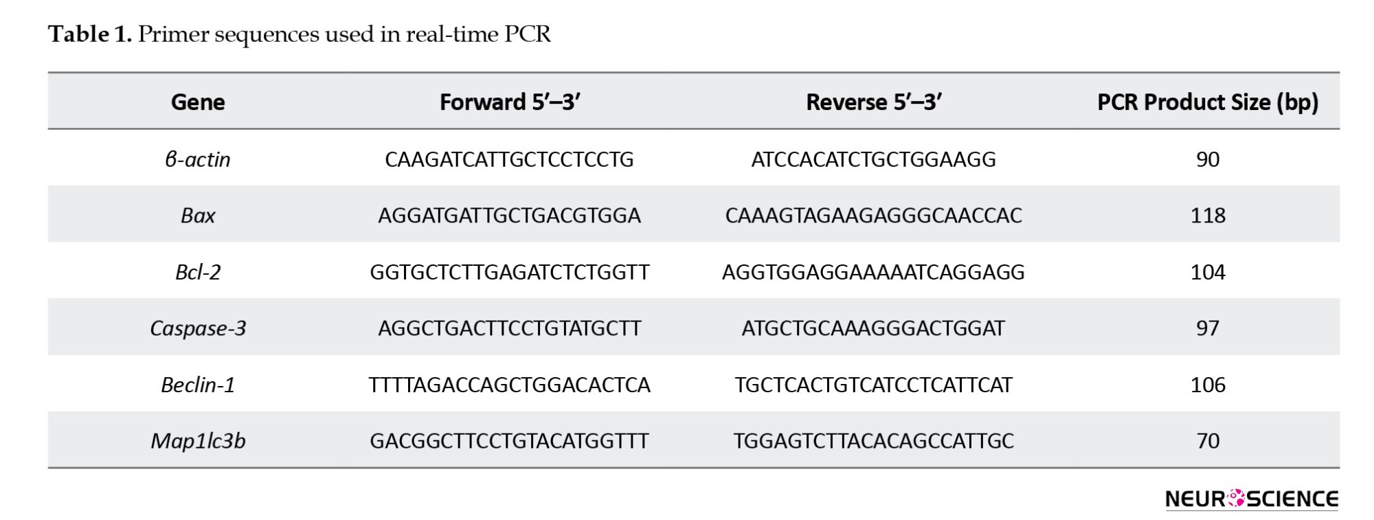
Expression levels were normalized to β-actin, and the 2–ΔΔCt method was used to calculate relative expression levels (Gholipour et al., 2023; Mirsanei et al., 2023). Primer sequences are provided in Table 1.
Histological analysis and TUNEL assay
After fixation, cerebellar tissues were embedded in paraffin, sectioned at 5 µm thickness using a microtome (Didsabz DS8402, Iran), and stained with cresyl violet (Nissl stain). For each animal, 20 coronal cerebellar sections were analyzed. The entire Purkinje cell layer was evaluated for neuronal counts in a blinded manner (Akhavan Tavakoli et al., 2023; Bakhtiary et al., 2010).
For apoptosis detection, paraffin-embedded sections underwent deparaffinization and rehydration followed by the TUNEL assay using a commercial kit (Roche, Germany). The procedure involved:
• PBS wash (20 min, room temperature)
• Peroxidase blocking (3% H₂O₂ in methanol, 10 min)
• Permeabilization (sodium citrate/Triton X-100 solution, 15 min, 2–8 °C)
• Incubation with TUNEL reaction mixture (1 h, room temperature)
Propidium iodide (PI) was used for nuclear counterstaining. TUNEL-positive cells were visualized under a fluorescence microscope (Olympus, Tokyo, Japan) with a green filter at 40× magnification (Nazari et al., 2023; Pourheydar et al., 2016).
Statistical analysis
All data analyses were conducted using GraphPad Prism software, version 8. The normality of data distribution was assessed using the Kolmogorov–Smirnov test. Group comparisons and detection of significant differences between variables were performed using one-way ANOVA for gene expression data and two-way ANOVA for histological analysis. Tukey’s post hoc test was subsequently applied to identify specific group differences. Data are presented as Mean±SD, with a P<0.05 considered indicative of statistical significance.
Results
Characterization of nanostructures
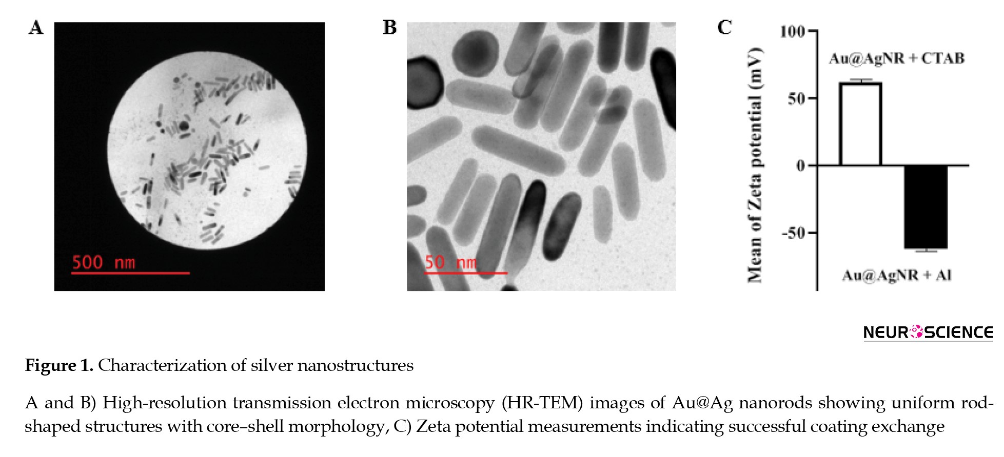
HR-TEM revealed that the synthesized Au@Ag nanostructures were rod-shaped with a uniform morphology and core–shell architecture (Figures 1A and 1B).
Metal-based nanoparticles, especially silver nanoparticles (AgNPs), have attracted significant interest in nanomedicine due to their unique physicochemical and biological properties, such as antimicrobial, anticancer, and catalytic activities (Sánchez-López et al., 2020; Sofi et al., 2021). AgNPs are widely incorporated into commercial and medical products, including wound dressings, cosmetics, food packaging, dental materials, and drug delivery systems (Ahmed et al., 2017; Thakur et al., 2014). However, growing evidence indicates that AgNPs pose potential health risks, as exposure through inhalation, ingestion, dermal absorption, or intravenous routes can lead to their accumulation in organs, like the liver, spleen, kidneys, and brain (Ahmed et al., 2017; Demir, 2021; Wei et al., 2015). Their toxicity is primarily linked to oxidative stress, ionic silver (Ag+) release, mitochondrial dysfunction, and apoptosis induction (Demir, 2021; Dos Santos et al., 2014). Anisotropic AgNPs, such as nanorods, display improved physical properties over spherical particles; however, their biocompatibility requires thorough evaluation (Durairaj et al., 2019). Coating strategies using polymers, like sodium alginate can enhance stability and minimize toxicity by modulating surface reactivity (Demir, 2021).
Critically, AgNPs can penetrate the blood–brain barrier (BBB) through passive diffusion or endocytic pathways, leading to accumulation within the central nervous system (CNS) (Ghooshchian et al., 2017; Hoet et al., 2004; Trickler et al., 2010). AgNP-induced neurotoxicity has been attributed to inflammation, BBB disruption, and direct damage to neurons and glial cells. The extent of toxicity varies with particle size, surface coating, and dosage (Mohamed et al., 2021). Previous in vivo studies in rodents have demonstrated cognitive impairments and synaptic alterations following AgNP exposure, with mechanisms implicating oxidative stress, altered gene expression, and apoptosis (Ahamed et al., 2010; Nejati et al., 2018; Yin et al., 2013). Given their ability to bypass natural biological barriers and exert neurotoxic effects, a comprehensive understanding of AgNPs’ long-term impact on neural tissues is critical.
Although anisotropic AgNPs have better properties than spherical ones, it is crucial to assess the biocompatibility and toxicity of these anisotropic AgNPs (Durairaj et al., 2019). Despite numerous studies on acute exposure and spherical nanoparticles, the chronic effects of anisotropic AgNPs and their underlying molecular mechanisms remain less well understood. This study investigated the acute and chronic toxicity of a novel AgNP structure—alginate-coated Au@Ag core–shell nanorods—on the mouse cerebellum, evaluating apoptotic and autophagic markers alongside histological changes to elucidate their impact on cerebellar integrity and function.
Materials and Methods
Synthesis and characterization of silver nanostructures
As previously described (Nazari et al., 2023), gold-silver core–shell nanorods (Au@Ag) were synthesized using a seed-mediated growth method. The nanostructures were subsequently characterized to confirm morphology, size, and core–shell composition using high-resolution transmission electron microscopy (HR-TEM).
Animal groups and experimental design
Forty adult male mice (5 weeks old, 25–30 g) were obtained from the Center of Experimental and Comparative Studies at Iran University of Medical Sciences. All experimental protocols adhered to the guidelines set by the National Institutes of Health (NIH) and were approved by the Institutional Animal Ethics Committee at Iran University of Medical Sciences, Tehran, Iran.
Mice were housed under standard conditions: a 12-hour light/dark cycle at a temperature of 22–25 °C with unrestricted access to food and water. Animals were randomly assigned to five experimental groups (n=8 per group):
Control group: No treatment.
Sham group 1 (Al-14): Received alginate solution (1.5 mg/kg/day, intraperitoneally) (Nazari et al., 2023) for 2 weeks (Xu et al., 2015).
Sham group 2 (Al-35): Received alginate solution (1.5 mg/kg/day, intraperitoneally) for 5 weeks.
Treatment group 1 (Ag-14): Received Ag nanostructures (1.5 mg/kg/day, intraperitoneally) for 2 weeks.
Treatment group 2 (Ag-35): Received Ag nanostructures (1.5 mg/kg/day, intraperitoneally) for 5 weeks.
At the end of the treatment period, mice were deeply anesthetized using ketamine (20 mg/kg) and xylazine (0.64 mg/kg). Cardiac perfusion was performed by inserting a needle into the left ventricle and flushing with physiological saline followed by 4% paraformaldehyde to fix the tissues. The right atrium was incised to allow exsanguination. After perfusion, the head was removed and post-fixed in 4% paraformaldehyde for 24 hours. The brain was then dissected and the cerebellar tissue was separated from the whole brain. A portion was stored at −80 °C for quantitative real-time PCR analysis (n=3 of each group), while the remaining tissue was reserved in 10% paraformaldehyde for subsequent histological processing (n=2 for TUNEL assay and n=2 for histological analysis).
RNA isolation, cDNA synthesis, and quantitative real-time PCR
Total RNA was extracted from cerebellar tissues using TRIzol reagent (Millipore Sigma, USA) and chloroform, following the manufacturer’s protocol. RNA concentration and purity were assessed with a NanoDrop spectrophotometer (Thermo Fisher Scientific, USA) using absorbance ratios at 260/280 nm.
First-strand complementary DNA (cDNA) was synthesized using a commercial cDNA synthesis kit (Fermentas, Thermo Fisher Scientific, USA). Quantitative real-time PCR (qRT-PCR) was performed using a Corbett Research qPCR system (Australia) to analyze gene expression of autophagy markers (Beclin-1 and LC3) and apoptosis markers (Bax, Bcl-2, and Caspase-3).
The 10 µL qRT-PCR reaction mixture included:
• 5 pmol of specific forward and reverse primers
• 250 ng of cDNA template
• SYBR Green/ROX dye solution (25:1 ratio)
Expression levels were normalized to β-actin, and the 2–ΔΔCt method was used to calculate relative expression levels (Gholipour et al., 2023; Mirsanei et al., 2023). Primer sequences are provided in Table 1.
Histological analysis and TUNEL assay
After fixation, cerebellar tissues were embedded in paraffin, sectioned at 5 µm thickness using a microtome (Didsabz DS8402, Iran), and stained with cresyl violet (Nissl stain). For each animal, 20 coronal cerebellar sections were analyzed. The entire Purkinje cell layer was evaluated for neuronal counts in a blinded manner (Akhavan Tavakoli et al., 2023; Bakhtiary et al., 2010).
For apoptosis detection, paraffin-embedded sections underwent deparaffinization and rehydration followed by the TUNEL assay using a commercial kit (Roche, Germany). The procedure involved:
• PBS wash (20 min, room temperature)
• Peroxidase blocking (3% H₂O₂ in methanol, 10 min)
• Permeabilization (sodium citrate/Triton X-100 solution, 15 min, 2–8 °C)
• Incubation with TUNEL reaction mixture (1 h, room temperature)
Propidium iodide (PI) was used for nuclear counterstaining. TUNEL-positive cells were visualized under a fluorescence microscope (Olympus, Tokyo, Japan) with a green filter at 40× magnification (Nazari et al., 2023; Pourheydar et al., 2016).
Statistical analysis
All data analyses were conducted using GraphPad Prism software, version 8. The normality of data distribution was assessed using the Kolmogorov–Smirnov test. Group comparisons and detection of significant differences between variables were performed using one-way ANOVA for gene expression data and two-way ANOVA for histological analysis. Tukey’s post hoc test was subsequently applied to identify specific group differences. Data are presented as Mean±SD, with a P<0.05 considered indicative of statistical significance.
Results
Characterization of nanostructures
HR-TEM revealed that the synthesized Au@Ag nanostructures were rod-shaped with a uniform morphology and core–shell architecture (Figures 1A and 1B).
The gold nanorod core was coated with a silver shell approximately 2.8 nm thick. The average length of the nanorods was 50–60 nm with a diameter of ~25 nm, yielding an aspect ratio of approximately 2.4. Zeta potential analysis demonstrated a substantial surface charge shift following alginate coating, with values of +62.15±1.91 mV for CTAB-coated and −62.49±1.29 mV for alginate-coated nanostructures, confirming successful surface modification (Figure 1C).
Gene expression in cerebellar tissue
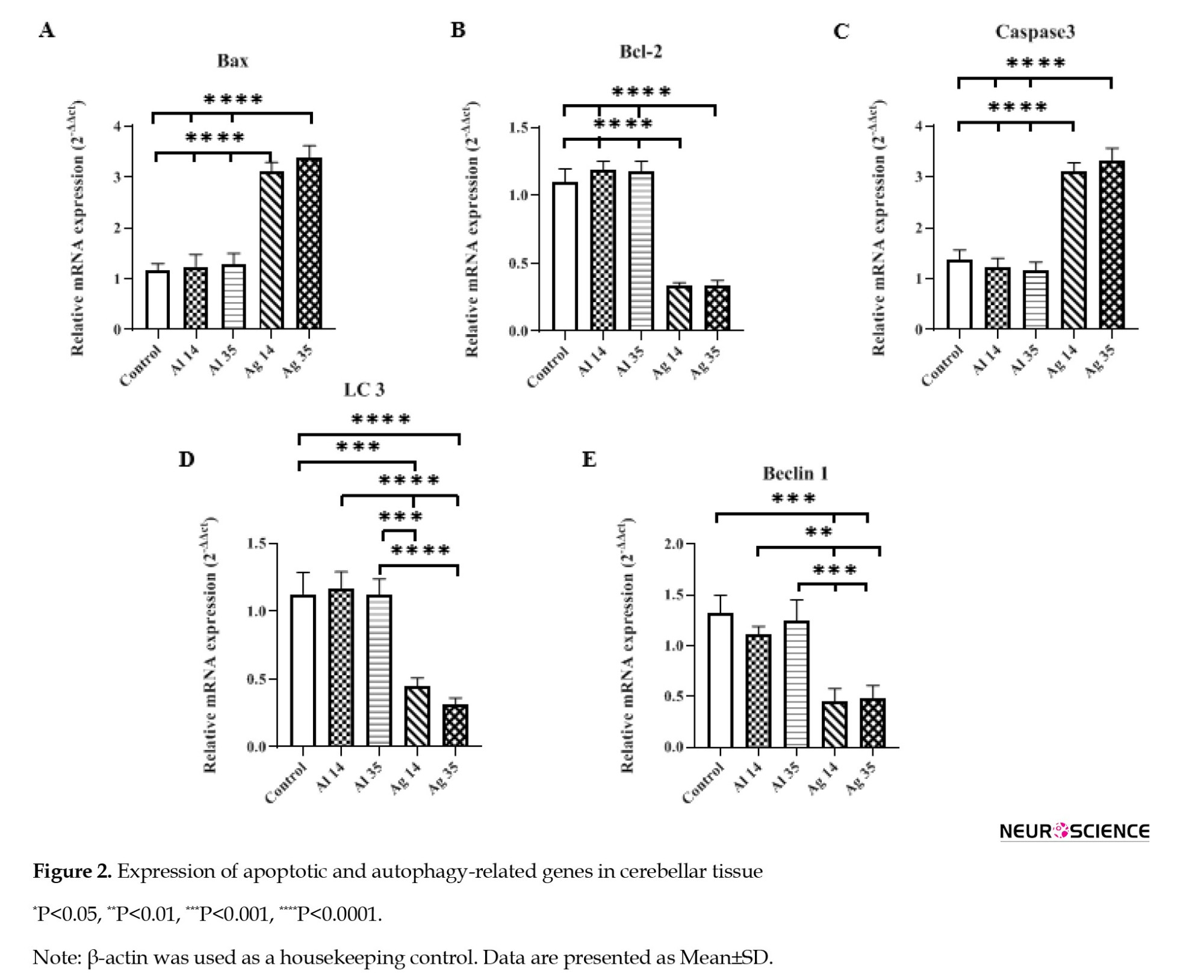
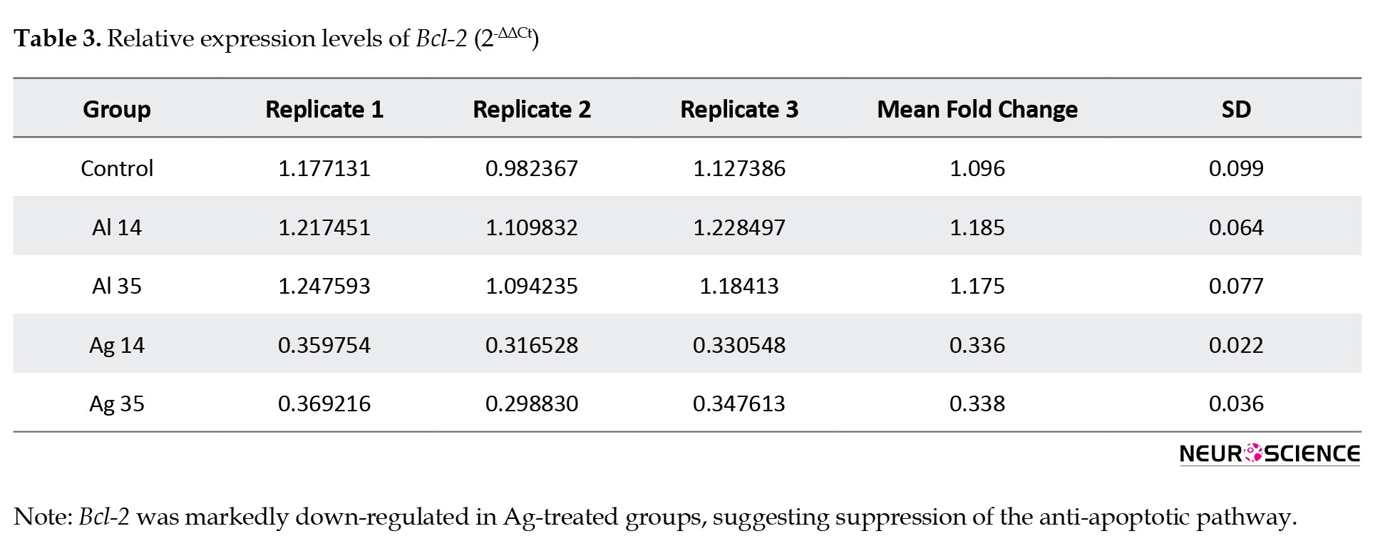
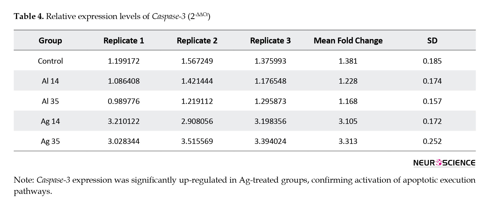
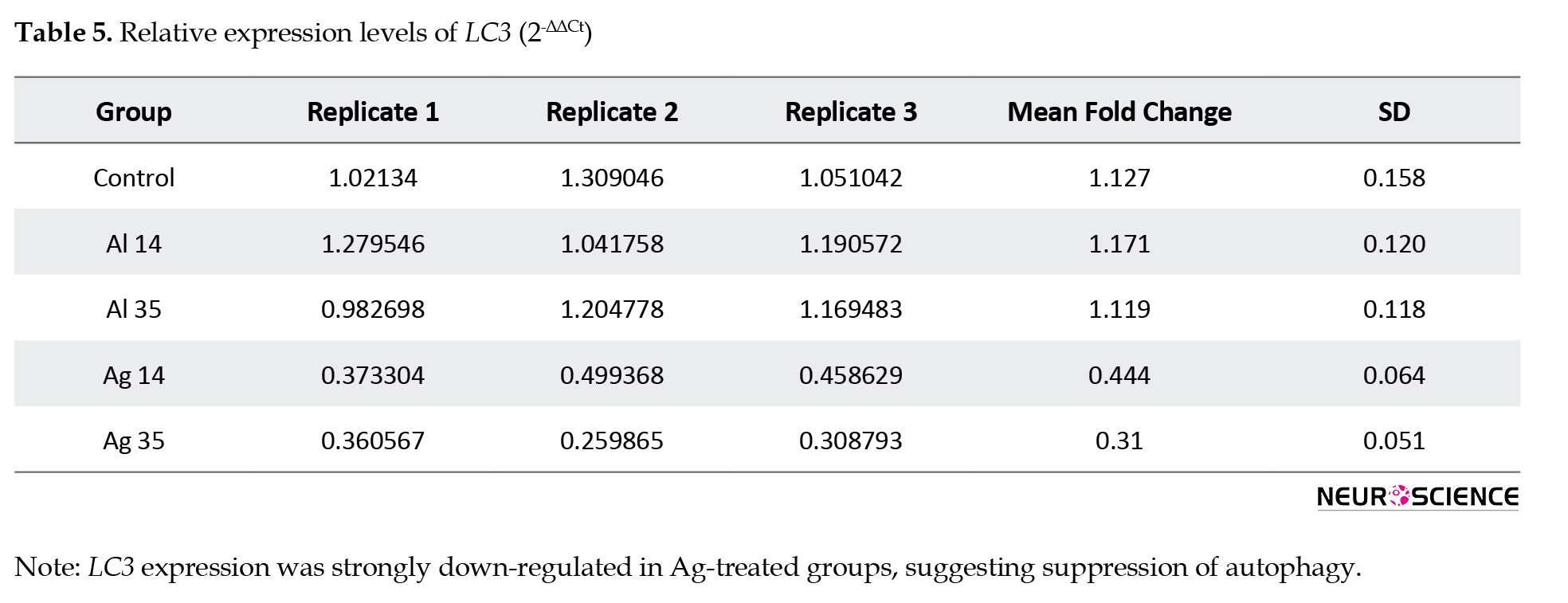
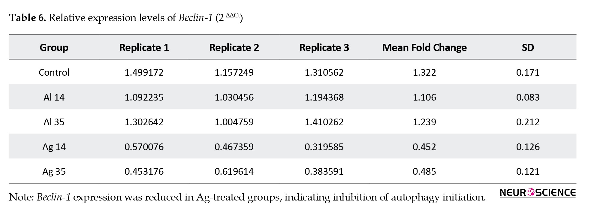
Quantitative RT-PCR analysis revealed significant alterations in the expression of apoptosis- and autophagy-related genes in the cerebellum of mice treated with silver nanostructures (Figure 2).
Gene expression in cerebellar tissue
Quantitative RT-PCR analysis revealed significant alterations in the expression of apoptosis- and autophagy-related genes in the cerebellum of mice treated with silver nanostructures (Figure 2).
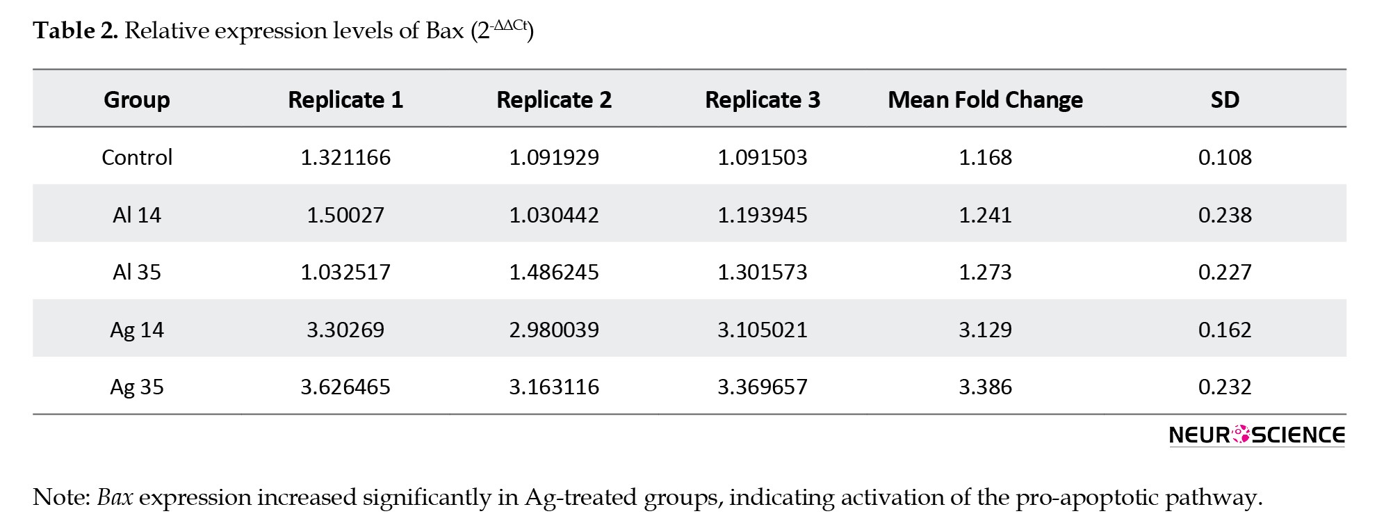
Expression of Bax and Caspase-3 was significantly upregulated in both Ag 14 and Ag 35 groups compared to control and sham groups, indicating enhanced apoptotic signaling. Conversely, expression of the anti-apoptotic gene Bcl-2 was significantly downregulated in the Ag-treated groups. Expression of LC3, an autophagic marker, was reduced in the treatment groups compared to the control (P=0.0001 and P<0.0001) and sham (P=0.0002 and P<0.0001) groups. Similarly, Beclin-1 also showed downregulation in the Ag 14 and Ag 35 groups compared to the control (control vs Ag 14: P=0.0002; control vs Ag 35: P=0.0003) and sham (Al 14 vs Ag 14: P=0.0022; Al 14 vs Ag 35: P=0.0033; Al 35 vs Ag 14: P=0.0005; Al 35 vs Ag 35: P=0.0007) groups. These results suggest suppression of autophagic activity in cerebellar tissue exposed to silver nanostructures. Tables 2, 3, 4, 5, and 6 present the fold-change values obtained from the qPCR analysis.
Histological analysis and TUNEL assay in cerebellar tissue
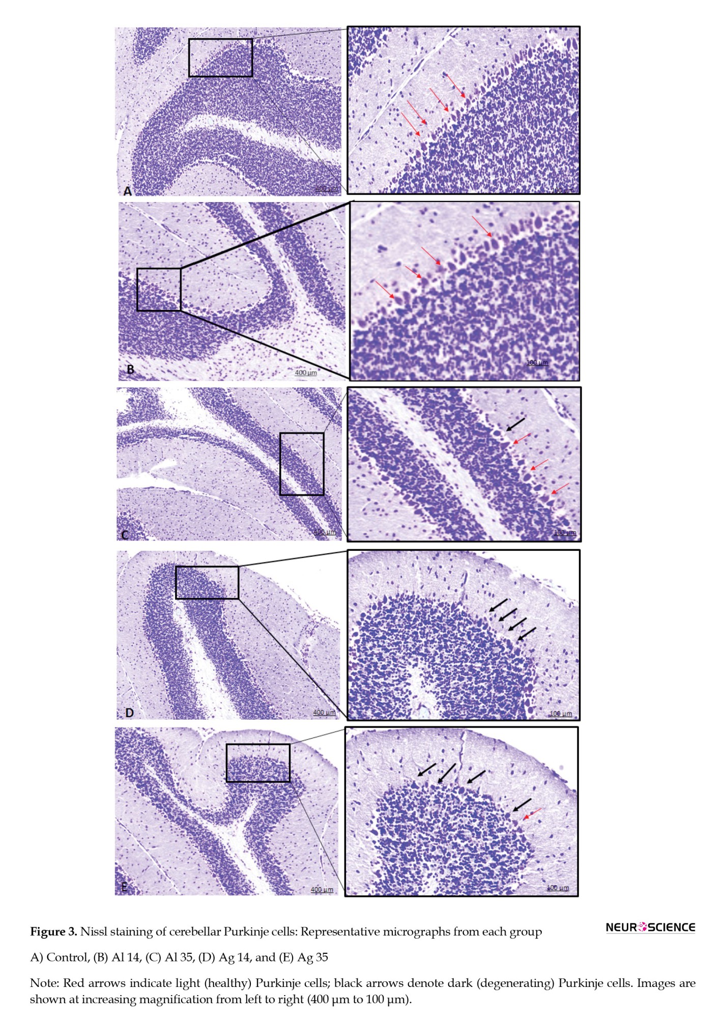
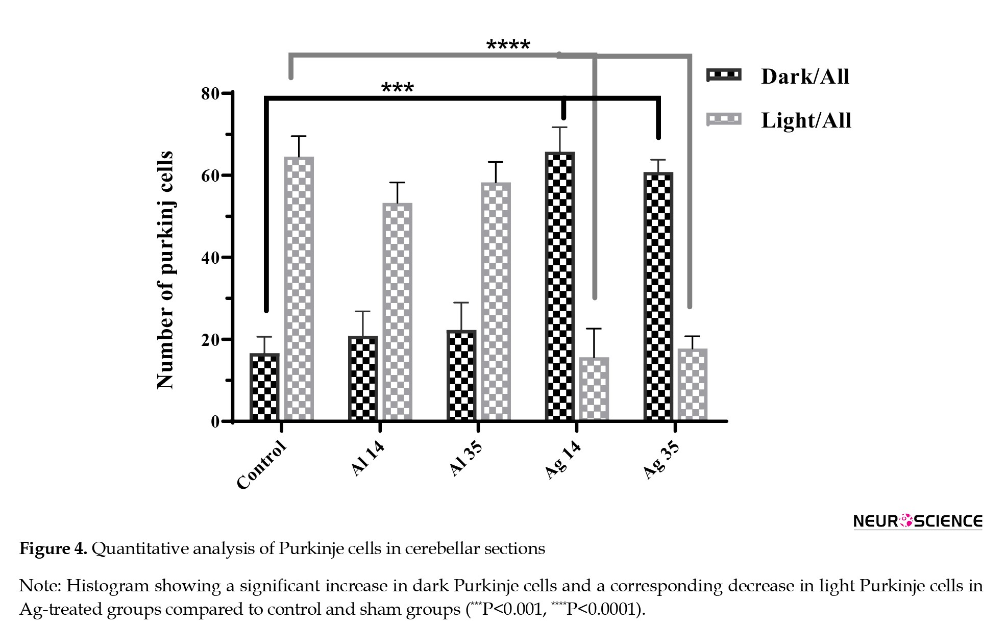
Cresyl violet (Nissl) staining identified two distinct Purkinje cell types: light cells with clearly defined nuclei and cytoplasm, representing healthy neurons, and dark cells, characterized by shrunken, basophilic cytoplasm and indistinct nuclear features, consistent with neuronal injury or degeneration (Hassanzadeh et al., 2018). A marked increase in the number of dark Purkinje cells was observed in Ag-treated groups compared to control and sham groups (P<0.0001), while the number of light cells was significantly decreased (Figures 3 and 4).
Histological analysis and TUNEL assay in cerebellar tissue
Cresyl violet (Nissl) staining identified two distinct Purkinje cell types: light cells with clearly defined nuclei and cytoplasm, representing healthy neurons, and dark cells, characterized by shrunken, basophilic cytoplasm and indistinct nuclear features, consistent with neuronal injury or degeneration (Hassanzadeh et al., 2018). A marked increase in the number of dark Purkinje cells was observed in Ag-treated groups compared to control and sham groups (P<0.0001), while the number of light cells was significantly decreased (Figures 3 and 4).
Quantitative analysis revealed that the number of dark Purkinje cells significantly increased in Ag 14 (49.08±5.2) and Ag 35 (44.17±5.2) groups compared to controls. Conversely, light cell counts were significantly reduced in both treatment groups (P<0.0001).
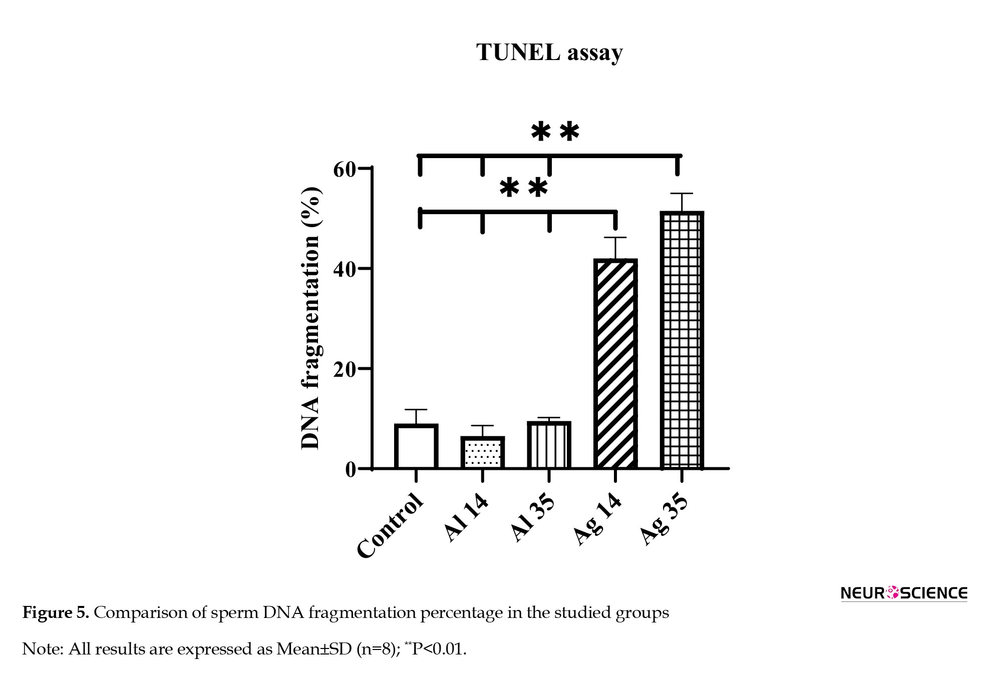
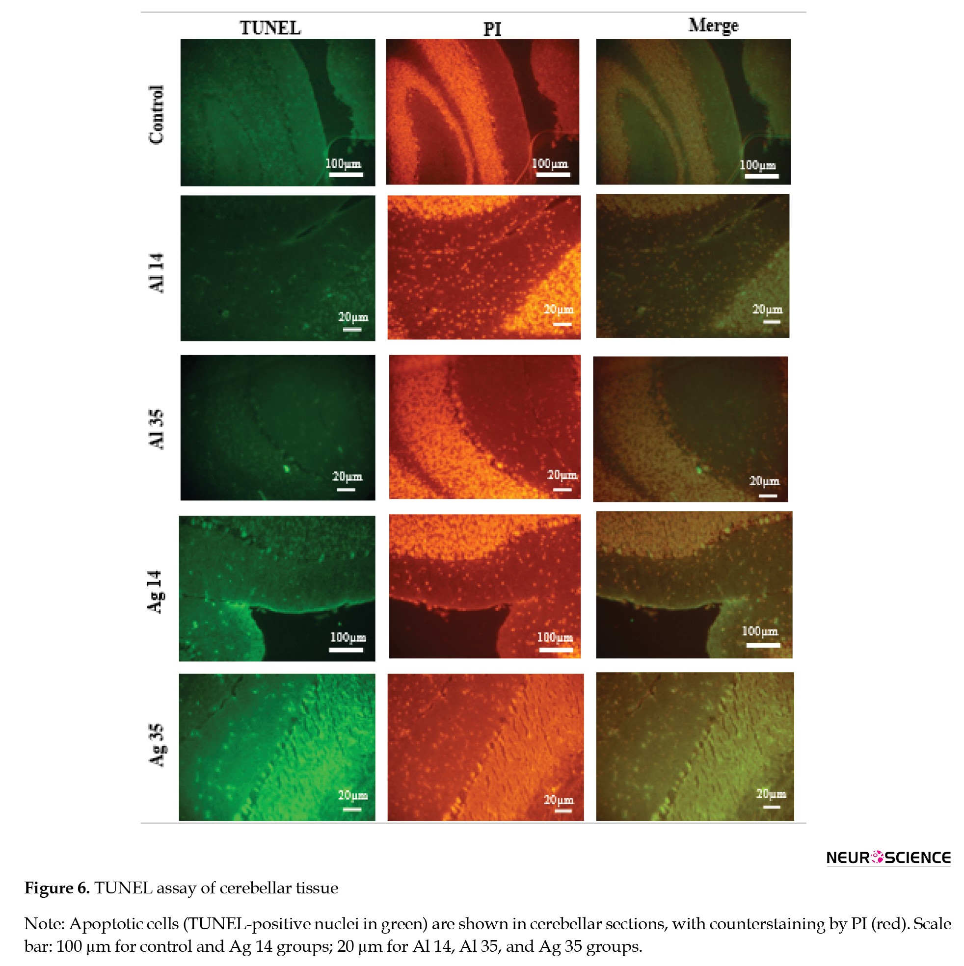
To further confirm apoptosis, TUNEL staining was performed. Quantitative evaluation of the results showed a significant increase in TUNEL-positive nuclei (green fluorescence) in the Ag 14 (P=0.0050) and especially the Ag 35 (P=0.0019) groups, consistent with apoptotic gene expression and histopathological findings (Figures 5 and 6).
To further confirm apoptosis, TUNEL staining was performed. Quantitative evaluation of the results showed a significant increase in TUNEL-positive nuclei (green fluorescence) in the Ag 14 (P=0.0050) and especially the Ag 35 (P=0.0019) groups, consistent with apoptotic gene expression and histopathological findings (Figures 5 and 6).
Nuclei were counterstained with propidium iodide (PI, red) to facilitate visualization.
Discussion
This study explored both the acute and chronic effects of alginate-coated Au@Ag core–shell nanorods on cerebellar tissue, with particular attention to their influence on apoptotic and autophagic pathways. Treatment with these nanostructures led to a significant upregulation of pro-apoptotic genes, such as Caspase-3 and Bax, along with a marked reduction in the expression of the anti-apoptotic gene Bcl-2. In parallel, we observed a downregulation of autophagy-related markers, LC3 and Beclin-1, in the cerebellum of treated animals. Histological evaluation further showed an increased presence of dark Purkinje cells, a recognized indicator of neuronal injury, while TUNEL staining confirmed an increase in DNA fragmentation and apoptotic signals, particularly in the chronically exposed group. Together, these results suggest that Au@Ag nanorods disrupt cerebellar cellular homeostasis by simultaneously promoting apoptosis and impairing autophagy.
Silver is widely valued for its antimicrobial activity and high conductivity and, in its bulk form, is generally regarded as safe. However, in its ionic form (Ag⁺), it becomes highly toxic, with documented effects on organisms across multiple biological systems, even at microgram-per-liter concentrations (Mertens et al., 2019). AgNPs, owing to their unique surface area and reactivity, have become prominent in various technological and medical applications. Yet, their potential for toxicity remains a critical concern. Several factors influence AgNP toxicity, including particle size, chemical composition, surface coating, concentration, duration and route of exposure, and especially their capacity to generate oxidative stress, which is widely regarded as a central mechanism of nanoparticle-induced cellular damage (Gromadzka-Ostrowska et al., 2012).
Bimetallic nanostructures, particularly those combining gold and silver in core–shell configurations, have demonstrated functional advantages, such as enhanced optical properties and biocompatibility. Gold offers structural stability and biological inertness, while silver enhances sensitivity in applications, like surface-enhanced Raman scattering (SERS) (Kvitek et al., 2020). Despite these benefits, the neurotoxicity profile of these nanostructures—especially when coated with biocompatible materials, like alginate—remains poorly understood. This study is, to our knowledge, the first to evaluate the cerebellar impact of such modified Au@Ag nanorods, revealing that even with an alginate coating, significant adverse effects on neuronal integrity can occur.
Apoptosis and autophagy are essential biological processes that preserve tissue homeostasis and ensure the removal of damaged or unnecessary cells. They are tightly regulated and often interact with each other, sharing molecular regulators and signaling cascades (Nikoletopoulou et al., 2013). Apoptosis plays a key role in development and immune function, and its regulation is critically governed by proteins of the Bcl-2 family (Honardoost et al., 2013; Macchi et al., 2015). These proteins include both pro-apoptotic members, such as Bax and Bak, and anti-apoptotic counterparts, like Bcl-2 and Bcl-xL (Hassan et al., 2014). Bcl-2 can prevent the release of cytochrome c from mitochondria, thus inhibiting caspase activation and cell death. Meanwhile, Beclin-1, a key autophagy regulator, is known to form a complex with Bcl-2, which can inhibit autophagic activity. During apoptosis, Beclin-1 and other autophagy-related proteins, such as Atg3 may be cleaved by caspases, linking the two processes through shared degradation pathways (Nikoletopoulou et al., 2013; Oral et al., 2012). A shift in the Bax/Bcl-2 ratio in favor of apoptosis, as seen in our study, not only promotes programmed cell death but may also suppress autophagic processes by disrupting these regulatory complexes (Aguzzi & O’connor, 2010).
The increase in dark Purkinje cells observed in Nissl-stained sections supports this conclusion, indicating structural deterioration commonly associated with oxidative or toxic stress. Additionally, the strong TUNEL positivity in Ag-treated groups confirms that DNA fragmentation—an established hallmark of apoptosis—is a major outcome of nanoparticle exposure in this model. Taken together, the molecular, histological, and cytological data in this study provide converging evidence of cerebellar toxicity induced by prolonged exposure to alginate-coated Au@Ag nanostructures. To date, no comprehensive investigation has systematically examined the biodistribution of alginate-coated Au@Ag core–shell nanorods within cerebellar tissue, and current studies primarily focus on spherical particles and brain regions, especially the hippocampus.
Janzadeh et al., in a meta-analysis, showed that AgNPs can induce neuronal death after crossing into the brain. Both AgNPs smaller and larger than 10 nm were capable of causing neuronal cell damage. This neurotoxic effect persisted for an extended period, lasting up to six months. Neurons from embryos of mothers exposed to AgNPs during pregnancy were similarly affected as those from animals directly exposed to the nanoparticles. Additionally, AgNP exposure was linked to impairments in memory and cognitive functions. Research suggests that inflammation and elevated oxidative stress, leading to apoptosis, are the primary mechanisms driving AgNP-induced toxicity (Janzadeh et al., 2022).
Consistent with our findings, Yin et al. conducted an in-depth investigation into the neurotoxic effects of commercially available AgNPs on rat cerebellar granule cells (CGCs) and elucidated the underlying molecular mechanisms. Their study demonstrated that AgNPs significantly compromise the viability of primary neuronal cells by inducing apoptosis associated with oxidative stress, mediated through caspase-dependent signaling pathways (Yin et al., 2013). In another study, Mohamed et al. explored the impact of AgNPs on the rat cerebellar cortex. Their findings demonstrated that exposure to Ag-NPs induced biochemical, cellular, and molecular alterations in the cerebellar cortex in a dose-dependent fashion. The primary mechanisms involved the activation of apoptotic pathways alongside increased oxidative stress and inflammation (Mohamed et al., 2020). Yin et al. also showed that exposure to AgNPs leads to cerebellar ataxia-like symptoms in rats, characterized by impaired motor coordination and reduced locomotor activity. Their results indicate that this motor dysfunction is linked to a decrease in CACNA1A expression, shedding light on the molecular mechanisms underlying AgNPs’ neurotoxic effects (Yin et al., 2015).
The widespread use of AgNPs in water purification, biosensors, nasal decongestants, oral hygiene products, long-term wound dressings, and various cosmetics today represents significant risk factors for human exposure to AgNPs (Mohamed et al., 2021). Accordingly, it is suggested to investigate other shapes, structures, biocompatible coatings, and techniques to reduce the unexpected harmful effects of AgNPs in biological applications.
Challenges and limitations
Specifically, while the TUNEL assay and gene expression analyses provide valuable insights into apoptosis and autophagy, they do not capture the full complexity of molecular pathways involved in nanostructure-induced neurotoxicity. Additionally, the study is limited to male mice, which may not fully represent sex-specific responses. Furthermore, the acute and chronic exposure durations chosen may not encompass all potential exposure scenarios relevant to humans. These limitations highlight the need for further in-depth studies employing complementary techniques and diverse models to fully elucidate the neurotoxic effects of alginate-coated silver nanostructures.
Conclusion
This study demonstrates that alginate-coated Au@Ag core–shell nanorods exert toxic effects on cerebellar tissue by activating apoptotic pathways and inhibiting autophagy, particularly with prolonged exposure. Despite the use of a biocompatible alginate coating, these nanostructures induced significant neuronal degeneration and molecular alterations in the cerebellum.
Given the increasing use of AgNPs in medical and commercial products, our findings underscore the importance of further research into their long-term safety. Future studies should focus on developing strategies to reduce nanoparticle toxicity—such as optimizing particle size, dose, and surface chemistry—to ensure safe integration of these materials into biomedical applications while minimizing potential risks to neural health and human safety.
Ethical Considerations
Compliance with ethical guidelines
This study was approved by the Research Ethics Committee of Iran University of Medical Sciences (IUMS), Tehran, Iran (Code: IR.IUMS.REC.1400.1069). The experimental methodology was strictly adhered to the requirements for the use of laboratory animals and the arrive guidelines.
Funding
The present study was supported by Iran University of Medical Sciences (IUMS), Tehran, Iran (Grant No.: 1400-3-20-22370).
Authors' contributions
Conceptualization: Mahsa Nazari; Methodology: Mahsa Nazari and Ronak Shabani; Project administration: Mehdi Mehdizadeh; Investigation: Mahsa Nazari, Sahar Eghbali and Maryam Akhavan Tavakoli; Formal analysis, data curation, and data interpretation: Rana Mehdizadeh; Writing the original draft: Mahsa Nazari and Maryam Akhavan Tavakoli; Review and editing: Ronak Shabani; Final approval: Sahar Hakimpour, Ronak Shabani, and Asghar Marzban; Supervision: Asghar Marzban.
Conflict of interest
The authors declared no conflict of interest.
Acknowledgments
The experiments described in this study were conducted within the laboratories of the Anatomy Department and Cellular and Molecular Research Center (CMRC) at Iran University of Medical Sciences (IUMS), Tehran, Iran. We would like to acknowledge the support provided by the Iran University of Medical Sciences in facilitating this research.
References
Aguzzi, A., & O'Connor, T. (2010). Protein aggregation diseases: pathogenicity and therapeutic perspectives. Nature reviews. Drug Discovery, 9(3), 237–248. [DOI:10.1038/nrd3050] [PMID]
Ahamed, M., Posgai, R., Gorey, T. J., Nielsen, M., Hussain, S. M., & Rowe, J. J. (2010). Silver nanoparticles induced heat shock protein 70, oxidative stress and apoptosis in Drosophila melanogaster. Toxicology and Applied Pharmacology, 242(3), 263–269. [DOI:10.1016/j.taap.2009.10.016] [PMID]
Ahmed, S. M., Abdelrahman, S. A., & Shalaby, S. M. (2017). Evaluating the effect of silver nanoparticles on testes of adult albino rats (histological, immunohistochemical and biochemical study). Journal of Molecular Histology, 48(1), 9-27. [DOI:10.1007/s10735-016-9701-4] [PMID]
Akhavan Tavakoli, M., Soleimani, M., Marzban, H., Shabani, R., Moradi, F., & Ajdary, M., et al. (2023). Autophagic molecular alterations in the mouse cerebellum experimental autoimmune encephalomyelitis model following treatment with cannabidiol and fluoxetine. Molecular Neurobiology, 60(4), 1797-1809. [DOI:10.1007/s12035-023-03204-2] [PMID]
Bakhtiary, M., Marzban, M., Mehdizadeh, M., Joghataei, M. T., Khoei, S., & Pirhajati Mahabadi, V., et al. (2010). Comparison of transplantation of bone marrow stromal cells (BMSC) and stem cell mobilization by granulocyte colony stimulating factor after traumatic brain injury in rat. Iranian Biomedical Journal, 14(4), 142–149. [PMID]
Demir, E. (2021). A review on nanotoxicity and nanogenotoxicity of different shapes of nanomaterials. Journal of Applied Toxicology, 41(1), 118-147. [DOI:10.1002/jat.4061] [PMID]
Dos Santos, C. A., Seckler, M. M., Ingle, A. P., Gupta, I., Galdiero, S., & Galdiero, M., et al. (2014). Silver nanoparticles: Therapeutical uses, toxicity, and safety issues. Journal of Pharmaceutical Sciences, 103(7), 1931-44. [DOI:10.1002/jps.24001] [PMID]
Durairaj, K., Roy, B., Chandrasekaran, N., Krishnan, S. P., & Mukherjee, A. (2020). Silver nanorods induced oxidative stress and chromosomal aberrations in the Allium cepa model. IET Nanobiotechnology, 14(2), 161–166. [DOI:10.1049/iet-nbt.2019.0224] [PMID]
Gholipour, H., Amjadi, F. S., Zandieh, Z., Mehdizadeh, M., Ajdary, M., & Delbandi, A. A., et al. (2023). Investigation of the Effect of Seminal Plasma Exosomes from the normal and oligoasthenoteratospermic males in the implantation process. Reports of Biochemistry & Molecular Biology, 12(2), 294-305. [DOI:10.61186/rbmb.12.2.294] [PMID]
Ghooshchian, M., Khodarahmi, P., & Tafvizi, F. (2017). Apoptosis-mediated neurotoxicity and altered gene expression induced by silver nanoparticles. Toxicology and Industrial Health, 33(10), 757–764. [DOI:10.1177/0748233717719195] [PMID]
Gromadzka-Ostrowska, J., Dziendzikowska, K., Lankoff, A., Dobrzyńska, M., Instanes, C., & Brunborg, G., et al. (2012). Silver nanoparticles effects on epididymal sperm in rats. Toxicology Letters, 214(3), 251-258. [DOI:10.1016/j.toxlet.2012.08.028] [PMID]
Hassan, M., Watari, H., AbuAlmaaty, A., Ohba, Y., & Sakuragi, N. (2014). Apoptosis and molecular targeting therapy in cancer. BioMed Research International, 2014, 150845. [DOI:10.1155/2014/150845] [PMID]
Hassanzadeh, S., Jameie, S. B., Mehdizadeh, M., Soleimani, M., Namjoo, Z., & Soleimani, M. (2018). FNDC5 expression in Purkinje neurons of adult male rats with acute spinal cord injury following treatment with methylprednisolone. Neuropeptides, 70, 16-25. [DOI:10.1016/j.npep.2018.05.002] [PMID]
Hoet, P. H., Brüske-Hohlfeld, I., & Salata, O. V. (2004). Nanoparticles-known and unknown health risks. Journal of Nanobiotechnology, 2(1), 12. [DOI:10.1186/1477-3155-2-12] [PMID]
Honardoost, M., Soleimanjahi, H., & Rajaei, F. (2013). [Apoptosis: Programmed cell death (Persian)]. Journal of Inflammatory Disease, 17(3), 48-57. [Link]
Janzadeh, A., Behroozi, Z., Saliminia, F., Janzadeh, N., Arzani, H., & Tanha, K., et al. (2022). Neurotoxicity of silver nanoparticles in the animal brain: A systematic review and meta-analysis. Forensic Toxicology, 40(1), 49-63. [DOI:10.1007/s11419-021-00589-4] [PMID]
Kvitek, O., Havelka, V., Vesely, M., Reznickova, A., & Svorcik, V. (2020). Preparation of alloyed and “core-shell” Au/Ag bimetallic nanostructures on glass substrate by solid state dewetting. Journal of Alloys and Compounds, 829, 154627. [DOI:10.1016/j.jallcom.2020.154627]
Macchi, B., Marino-Merlo, F., Nocentini, U., Pisani, V., & Cuzzocrea, S., Grelli, S., et al. (2015). Role of inflammation and apoptosis in multiple sclerosis: comparative analysis between the periphery and the central nervous system. Journal of Neuroimmunology, 287, 80-87. [DOI:10.1016/j.jneuroim.2015.08.016] [PMID]
Mertens, J., Oorts, K., Leverett, D., & Arijs, K. (2019). Effects of silver nitrate are a conservative estimate for the effects of silver nanoparticles on algae growth and Daphnia magna reproduction. Environmental Toxicology and Chemistry, 38(8), 1701-1713. [DOI:10.1002/etc.4463] [PMID]
Mirsanei, J. S., Nazari, M., Shabani, R., Govahi, A., Eghbali, S., Ajdary, M., & Mehdizadeh, R., et al. (2023). Does gold-silver core-shell nanostructure with alginate coating induce apoptosis in human lymphoblastic tumoral (Jurkat) cell line? Reports of Biochemistry & Molecular Biology, 12(2), 233-240. [DOI:10.61186/rbmb.12.2.233] [PMID]
Mohamed, E. M., Kattaia, A. A. A., Abdul-Maksoud, R. S., & Abd El-Baset, S. A. (2020). Cellular, molecular and biochemical impacts of silver nanoparticles on rat cerebellar cortex. Cells, 10(1), 7. [DOI:10.3390/cells10010007] [PMID]
Nazari, M., Shabani, R., Ajdary, M., Ashjari, M., Shirazi, R., & Govahi, A., et al. (2023). Effects of Au@ Ag core-shell nanostructure with alginate coating on male reproductive system in mice. Toxicology Reports, 10, 104-116. [DOI:10.1016/j.toxrep.2023.01.003] [PMID]
Nejati, M., Tameh, A. A., Vahidinia, Z., & Atlasi, M. A. (2018). Mesenchymal stem cells improve ischemic stroke injury by anti-inflammatory properties in rat model of middle cerebral artery occlusion. Iranian Red Crescent Medical Journal, 20(1), e55085. [DOI:10.5812/ircmj.55085]
Nikoletopoulou, V., Markaki, M., Palikaras, K., & Tavernarakis, N. (2013). Crosstalk between apoptosis, necrosis and autophagy. Biochimica et Biophysica Acta, 1833(12), 3448–3459. [DOI:10.1016/j.bbamcr.2013.06.001] [PMID]
Oral, O., Oz-Arslan, D., Itah, Z., Naghavi, A., Deveci, R., & Karacali, S., et al. (2012). Cleavage of Atg3 protein by caspase-8 regulates autophagy during receptor-activated cell death. Apoptosis: An International Journal on Programmed Cell Death, 17(8), 810–820. [DOI:10.1007/s10495-012-0735-0] [PMID]
Pourheydar, B., Soleimani Asl, S., Azimzadeh, M., Rezaei Moghadam, A., Marzban, A., & Mehdizadeh, M. (2016). Neuroprotective effects of bone marrow mesenchymal stem cells on bilateral common carotid arteries occlusion model of cerebral ischemia in rat. Behavioural Neurology, 2016, 2964712. [DOI:10.1155/2016/2964712] [PMID]
Sánchez-López, E., Gomes, D., Esteruelas, G., Bonilla, L., Lopez-Machado, A. L., & Galindo, R., et al. (2020). Metal-based nanoparticles as antimicrobial agents: An overview. Nanomaterials, 10(2), 292. [DOI:10.3390/nano10020292] [PMID]
Sofi, M. A., Sunitha, S., Sofi, M. A., Pasha, S. K., & Choi, D. (2022). An overview of antimicrobial and anticancer potential of silver nanoparticles. Journal of King Saud University-Science, 34(2), 101791. [Link]
Thakur, M., Gupta, H., Singh, D., Mohanty, I. R., Maheswari, U., & Vanage, G., et al. (2014). Histopathological and ultra structural effects of nanoparticles on rat testis following 90 days (Chronic study) of repeated oral administration. Journal of Nanobiotechnology, 12, 42. [DOI:10.1186/s12951-014-0042-8] [PMID]
Trickler, W. J., Lantz, S. M., Murdock, R. C., Schrand, A. M., Robinson, B. L., & Newport, G. D., et al. (2010). Silver nanoparticle induced blood-brain barrier inflammation and increased permeability in primary rat brain microvessel endothelial cells. Toxicological Sciences: An Official Journal of the Society of Toxicology, 118(1), 160–170. [DOI:10.1093/toxsci/kfq244] [PMID]
Wei, L., Lu, J., Xu, H., Patel, A., Chen, Z. S., & Chen, G. (2015). Silver nanoparticles: synthesis, properties, and therapeutic applications. Drug Discovery Today, 20(5), 595–601. [DOI:10.1016/j.drudis.2014.11.014] [PMID]
Xu, L., Shao, A., Zhao, Y., Wang, Z., Zhang, C., & Sun, Y., et al. (2015). Neurotoxicity of silver nanoparticles in rat brain after intragastric exposure. Journal of Nanoscience and Nanotechnology, 15(6), 4215–4223. [DOI:10.1166/jnn.2015.9612] [PMID]
Yin, N., Liu, Q., Liu, J., He, B., Cui, L., & Li, Z., et al. (2013). Silver nanoparticle exposure attenuates the viability of rat cerebellum granule cells through apoptosis coupled to oxidative stress. Small, 9(9‐10), 1831-1841. [DOI:10.1002/smll.201202732] [PMID]
Yin, N., Zhang, Y., Yun, Z., Liu, Q., Qu, G., & Zhou, Q., et al. (2015). Silver nanoparticle exposure induces rat motor dysfunction through decrease in expression of calcium channel protein in cerebellum. Toxicology Letters, 237(2), 112-120. [DOI:10.1016/j.toxlet.2015.06.007] [PMID]
Discussion
This study explored both the acute and chronic effects of alginate-coated Au@Ag core–shell nanorods on cerebellar tissue, with particular attention to their influence on apoptotic and autophagic pathways. Treatment with these nanostructures led to a significant upregulation of pro-apoptotic genes, such as Caspase-3 and Bax, along with a marked reduction in the expression of the anti-apoptotic gene Bcl-2. In parallel, we observed a downregulation of autophagy-related markers, LC3 and Beclin-1, in the cerebellum of treated animals. Histological evaluation further showed an increased presence of dark Purkinje cells, a recognized indicator of neuronal injury, while TUNEL staining confirmed an increase in DNA fragmentation and apoptotic signals, particularly in the chronically exposed group. Together, these results suggest that Au@Ag nanorods disrupt cerebellar cellular homeostasis by simultaneously promoting apoptosis and impairing autophagy.
Silver is widely valued for its antimicrobial activity and high conductivity and, in its bulk form, is generally regarded as safe. However, in its ionic form (Ag⁺), it becomes highly toxic, with documented effects on organisms across multiple biological systems, even at microgram-per-liter concentrations (Mertens et al., 2019). AgNPs, owing to their unique surface area and reactivity, have become prominent in various technological and medical applications. Yet, their potential for toxicity remains a critical concern. Several factors influence AgNP toxicity, including particle size, chemical composition, surface coating, concentration, duration and route of exposure, and especially their capacity to generate oxidative stress, which is widely regarded as a central mechanism of nanoparticle-induced cellular damage (Gromadzka-Ostrowska et al., 2012).
Bimetallic nanostructures, particularly those combining gold and silver in core–shell configurations, have demonstrated functional advantages, such as enhanced optical properties and biocompatibility. Gold offers structural stability and biological inertness, while silver enhances sensitivity in applications, like surface-enhanced Raman scattering (SERS) (Kvitek et al., 2020). Despite these benefits, the neurotoxicity profile of these nanostructures—especially when coated with biocompatible materials, like alginate—remains poorly understood. This study is, to our knowledge, the first to evaluate the cerebellar impact of such modified Au@Ag nanorods, revealing that even with an alginate coating, significant adverse effects on neuronal integrity can occur.
Apoptosis and autophagy are essential biological processes that preserve tissue homeostasis and ensure the removal of damaged or unnecessary cells. They are tightly regulated and often interact with each other, sharing molecular regulators and signaling cascades (Nikoletopoulou et al., 2013). Apoptosis plays a key role in development and immune function, and its regulation is critically governed by proteins of the Bcl-2 family (Honardoost et al., 2013; Macchi et al., 2015). These proteins include both pro-apoptotic members, such as Bax and Bak, and anti-apoptotic counterparts, like Bcl-2 and Bcl-xL (Hassan et al., 2014). Bcl-2 can prevent the release of cytochrome c from mitochondria, thus inhibiting caspase activation and cell death. Meanwhile, Beclin-1, a key autophagy regulator, is known to form a complex with Bcl-2, which can inhibit autophagic activity. During apoptosis, Beclin-1 and other autophagy-related proteins, such as Atg3 may be cleaved by caspases, linking the two processes through shared degradation pathways (Nikoletopoulou et al., 2013; Oral et al., 2012). A shift in the Bax/Bcl-2 ratio in favor of apoptosis, as seen in our study, not only promotes programmed cell death but may also suppress autophagic processes by disrupting these regulatory complexes (Aguzzi & O’connor, 2010).
The increase in dark Purkinje cells observed in Nissl-stained sections supports this conclusion, indicating structural deterioration commonly associated with oxidative or toxic stress. Additionally, the strong TUNEL positivity in Ag-treated groups confirms that DNA fragmentation—an established hallmark of apoptosis—is a major outcome of nanoparticle exposure in this model. Taken together, the molecular, histological, and cytological data in this study provide converging evidence of cerebellar toxicity induced by prolonged exposure to alginate-coated Au@Ag nanostructures. To date, no comprehensive investigation has systematically examined the biodistribution of alginate-coated Au@Ag core–shell nanorods within cerebellar tissue, and current studies primarily focus on spherical particles and brain regions, especially the hippocampus.
Janzadeh et al., in a meta-analysis, showed that AgNPs can induce neuronal death after crossing into the brain. Both AgNPs smaller and larger than 10 nm were capable of causing neuronal cell damage. This neurotoxic effect persisted for an extended period, lasting up to six months. Neurons from embryos of mothers exposed to AgNPs during pregnancy were similarly affected as those from animals directly exposed to the nanoparticles. Additionally, AgNP exposure was linked to impairments in memory and cognitive functions. Research suggests that inflammation and elevated oxidative stress, leading to apoptosis, are the primary mechanisms driving AgNP-induced toxicity (Janzadeh et al., 2022).
Consistent with our findings, Yin et al. conducted an in-depth investigation into the neurotoxic effects of commercially available AgNPs on rat cerebellar granule cells (CGCs) and elucidated the underlying molecular mechanisms. Their study demonstrated that AgNPs significantly compromise the viability of primary neuronal cells by inducing apoptosis associated with oxidative stress, mediated through caspase-dependent signaling pathways (Yin et al., 2013). In another study, Mohamed et al. explored the impact of AgNPs on the rat cerebellar cortex. Their findings demonstrated that exposure to Ag-NPs induced biochemical, cellular, and molecular alterations in the cerebellar cortex in a dose-dependent fashion. The primary mechanisms involved the activation of apoptotic pathways alongside increased oxidative stress and inflammation (Mohamed et al., 2020). Yin et al. also showed that exposure to AgNPs leads to cerebellar ataxia-like symptoms in rats, characterized by impaired motor coordination and reduced locomotor activity. Their results indicate that this motor dysfunction is linked to a decrease in CACNA1A expression, shedding light on the molecular mechanisms underlying AgNPs’ neurotoxic effects (Yin et al., 2015).
The widespread use of AgNPs in water purification, biosensors, nasal decongestants, oral hygiene products, long-term wound dressings, and various cosmetics today represents significant risk factors for human exposure to AgNPs (Mohamed et al., 2021). Accordingly, it is suggested to investigate other shapes, structures, biocompatible coatings, and techniques to reduce the unexpected harmful effects of AgNPs in biological applications.
Challenges and limitations
Specifically, while the TUNEL assay and gene expression analyses provide valuable insights into apoptosis and autophagy, they do not capture the full complexity of molecular pathways involved in nanostructure-induced neurotoxicity. Additionally, the study is limited to male mice, which may not fully represent sex-specific responses. Furthermore, the acute and chronic exposure durations chosen may not encompass all potential exposure scenarios relevant to humans. These limitations highlight the need for further in-depth studies employing complementary techniques and diverse models to fully elucidate the neurotoxic effects of alginate-coated silver nanostructures.
Conclusion
This study demonstrates that alginate-coated Au@Ag core–shell nanorods exert toxic effects on cerebellar tissue by activating apoptotic pathways and inhibiting autophagy, particularly with prolonged exposure. Despite the use of a biocompatible alginate coating, these nanostructures induced significant neuronal degeneration and molecular alterations in the cerebellum.
Given the increasing use of AgNPs in medical and commercial products, our findings underscore the importance of further research into their long-term safety. Future studies should focus on developing strategies to reduce nanoparticle toxicity—such as optimizing particle size, dose, and surface chemistry—to ensure safe integration of these materials into biomedical applications while minimizing potential risks to neural health and human safety.
Ethical Considerations
Compliance with ethical guidelines
This study was approved by the Research Ethics Committee of Iran University of Medical Sciences (IUMS), Tehran, Iran (Code: IR.IUMS.REC.1400.1069). The experimental methodology was strictly adhered to the requirements for the use of laboratory animals and the arrive guidelines.
Funding
The present study was supported by Iran University of Medical Sciences (IUMS), Tehran, Iran (Grant No.: 1400-3-20-22370).
Authors' contributions
Conceptualization: Mahsa Nazari; Methodology: Mahsa Nazari and Ronak Shabani; Project administration: Mehdi Mehdizadeh; Investigation: Mahsa Nazari, Sahar Eghbali and Maryam Akhavan Tavakoli; Formal analysis, data curation, and data interpretation: Rana Mehdizadeh; Writing the original draft: Mahsa Nazari and Maryam Akhavan Tavakoli; Review and editing: Ronak Shabani; Final approval: Sahar Hakimpour, Ronak Shabani, and Asghar Marzban; Supervision: Asghar Marzban.
Conflict of interest
The authors declared no conflict of interest.
Acknowledgments
The experiments described in this study were conducted within the laboratories of the Anatomy Department and Cellular and Molecular Research Center (CMRC) at Iran University of Medical Sciences (IUMS), Tehran, Iran. We would like to acknowledge the support provided by the Iran University of Medical Sciences in facilitating this research.
References
Aguzzi, A., & O'Connor, T. (2010). Protein aggregation diseases: pathogenicity and therapeutic perspectives. Nature reviews. Drug Discovery, 9(3), 237–248. [DOI:10.1038/nrd3050] [PMID]
Ahamed, M., Posgai, R., Gorey, T. J., Nielsen, M., Hussain, S. M., & Rowe, J. J. (2010). Silver nanoparticles induced heat shock protein 70, oxidative stress and apoptosis in Drosophila melanogaster. Toxicology and Applied Pharmacology, 242(3), 263–269. [DOI:10.1016/j.taap.2009.10.016] [PMID]
Ahmed, S. M., Abdelrahman, S. A., & Shalaby, S. M. (2017). Evaluating the effect of silver nanoparticles on testes of adult albino rats (histological, immunohistochemical and biochemical study). Journal of Molecular Histology, 48(1), 9-27. [DOI:10.1007/s10735-016-9701-4] [PMID]
Akhavan Tavakoli, M., Soleimani, M., Marzban, H., Shabani, R., Moradi, F., & Ajdary, M., et al. (2023). Autophagic molecular alterations in the mouse cerebellum experimental autoimmune encephalomyelitis model following treatment with cannabidiol and fluoxetine. Molecular Neurobiology, 60(4), 1797-1809. [DOI:10.1007/s12035-023-03204-2] [PMID]
Bakhtiary, M., Marzban, M., Mehdizadeh, M., Joghataei, M. T., Khoei, S., & Pirhajati Mahabadi, V., et al. (2010). Comparison of transplantation of bone marrow stromal cells (BMSC) and stem cell mobilization by granulocyte colony stimulating factor after traumatic brain injury in rat. Iranian Biomedical Journal, 14(4), 142–149. [PMID]
Demir, E. (2021). A review on nanotoxicity and nanogenotoxicity of different shapes of nanomaterials. Journal of Applied Toxicology, 41(1), 118-147. [DOI:10.1002/jat.4061] [PMID]
Dos Santos, C. A., Seckler, M. M., Ingle, A. P., Gupta, I., Galdiero, S., & Galdiero, M., et al. (2014). Silver nanoparticles: Therapeutical uses, toxicity, and safety issues. Journal of Pharmaceutical Sciences, 103(7), 1931-44. [DOI:10.1002/jps.24001] [PMID]
Durairaj, K., Roy, B., Chandrasekaran, N., Krishnan, S. P., & Mukherjee, A. (2020). Silver nanorods induced oxidative stress and chromosomal aberrations in the Allium cepa model. IET Nanobiotechnology, 14(2), 161–166. [DOI:10.1049/iet-nbt.2019.0224] [PMID]
Gholipour, H., Amjadi, F. S., Zandieh, Z., Mehdizadeh, M., Ajdary, M., & Delbandi, A. A., et al. (2023). Investigation of the Effect of Seminal Plasma Exosomes from the normal and oligoasthenoteratospermic males in the implantation process. Reports of Biochemistry & Molecular Biology, 12(2), 294-305. [DOI:10.61186/rbmb.12.2.294] [PMID]
Ghooshchian, M., Khodarahmi, P., & Tafvizi, F. (2017). Apoptosis-mediated neurotoxicity and altered gene expression induced by silver nanoparticles. Toxicology and Industrial Health, 33(10), 757–764. [DOI:10.1177/0748233717719195] [PMID]
Gromadzka-Ostrowska, J., Dziendzikowska, K., Lankoff, A., Dobrzyńska, M., Instanes, C., & Brunborg, G., et al. (2012). Silver nanoparticles effects on epididymal sperm in rats. Toxicology Letters, 214(3), 251-258. [DOI:10.1016/j.toxlet.2012.08.028] [PMID]
Hassan, M., Watari, H., AbuAlmaaty, A., Ohba, Y., & Sakuragi, N. (2014). Apoptosis and molecular targeting therapy in cancer. BioMed Research International, 2014, 150845. [DOI:10.1155/2014/150845] [PMID]
Hassanzadeh, S., Jameie, S. B., Mehdizadeh, M., Soleimani, M., Namjoo, Z., & Soleimani, M. (2018). FNDC5 expression in Purkinje neurons of adult male rats with acute spinal cord injury following treatment with methylprednisolone. Neuropeptides, 70, 16-25. [DOI:10.1016/j.npep.2018.05.002] [PMID]
Hoet, P. H., Brüske-Hohlfeld, I., & Salata, O. V. (2004). Nanoparticles-known and unknown health risks. Journal of Nanobiotechnology, 2(1), 12. [DOI:10.1186/1477-3155-2-12] [PMID]
Honardoost, M., Soleimanjahi, H., & Rajaei, F. (2013). [Apoptosis: Programmed cell death (Persian)]. Journal of Inflammatory Disease, 17(3), 48-57. [Link]
Janzadeh, A., Behroozi, Z., Saliminia, F., Janzadeh, N., Arzani, H., & Tanha, K., et al. (2022). Neurotoxicity of silver nanoparticles in the animal brain: A systematic review and meta-analysis. Forensic Toxicology, 40(1), 49-63. [DOI:10.1007/s11419-021-00589-4] [PMID]
Kvitek, O., Havelka, V., Vesely, M., Reznickova, A., & Svorcik, V. (2020). Preparation of alloyed and “core-shell” Au/Ag bimetallic nanostructures on glass substrate by solid state dewetting. Journal of Alloys and Compounds, 829, 154627. [DOI:10.1016/j.jallcom.2020.154627]
Macchi, B., Marino-Merlo, F., Nocentini, U., Pisani, V., & Cuzzocrea, S., Grelli, S., et al. (2015). Role of inflammation and apoptosis in multiple sclerosis: comparative analysis between the periphery and the central nervous system. Journal of Neuroimmunology, 287, 80-87. [DOI:10.1016/j.jneuroim.2015.08.016] [PMID]
Mertens, J., Oorts, K., Leverett, D., & Arijs, K. (2019). Effects of silver nitrate are a conservative estimate for the effects of silver nanoparticles on algae growth and Daphnia magna reproduction. Environmental Toxicology and Chemistry, 38(8), 1701-1713. [DOI:10.1002/etc.4463] [PMID]
Mirsanei, J. S., Nazari, M., Shabani, R., Govahi, A., Eghbali, S., Ajdary, M., & Mehdizadeh, R., et al. (2023). Does gold-silver core-shell nanostructure with alginate coating induce apoptosis in human lymphoblastic tumoral (Jurkat) cell line? Reports of Biochemistry & Molecular Biology, 12(2), 233-240. [DOI:10.61186/rbmb.12.2.233] [PMID]
Mohamed, E. M., Kattaia, A. A. A., Abdul-Maksoud, R. S., & Abd El-Baset, S. A. (2020). Cellular, molecular and biochemical impacts of silver nanoparticles on rat cerebellar cortex. Cells, 10(1), 7. [DOI:10.3390/cells10010007] [PMID]
Nazari, M., Shabani, R., Ajdary, M., Ashjari, M., Shirazi, R., & Govahi, A., et al. (2023). Effects of Au@ Ag core-shell nanostructure with alginate coating on male reproductive system in mice. Toxicology Reports, 10, 104-116. [DOI:10.1016/j.toxrep.2023.01.003] [PMID]
Nejati, M., Tameh, A. A., Vahidinia, Z., & Atlasi, M. A. (2018). Mesenchymal stem cells improve ischemic stroke injury by anti-inflammatory properties in rat model of middle cerebral artery occlusion. Iranian Red Crescent Medical Journal, 20(1), e55085. [DOI:10.5812/ircmj.55085]
Nikoletopoulou, V., Markaki, M., Palikaras, K., & Tavernarakis, N. (2013). Crosstalk between apoptosis, necrosis and autophagy. Biochimica et Biophysica Acta, 1833(12), 3448–3459. [DOI:10.1016/j.bbamcr.2013.06.001] [PMID]
Oral, O., Oz-Arslan, D., Itah, Z., Naghavi, A., Deveci, R., & Karacali, S., et al. (2012). Cleavage of Atg3 protein by caspase-8 regulates autophagy during receptor-activated cell death. Apoptosis: An International Journal on Programmed Cell Death, 17(8), 810–820. [DOI:10.1007/s10495-012-0735-0] [PMID]
Pourheydar, B., Soleimani Asl, S., Azimzadeh, M., Rezaei Moghadam, A., Marzban, A., & Mehdizadeh, M. (2016). Neuroprotective effects of bone marrow mesenchymal stem cells on bilateral common carotid arteries occlusion model of cerebral ischemia in rat. Behavioural Neurology, 2016, 2964712. [DOI:10.1155/2016/2964712] [PMID]
Sánchez-López, E., Gomes, D., Esteruelas, G., Bonilla, L., Lopez-Machado, A. L., & Galindo, R., et al. (2020). Metal-based nanoparticles as antimicrobial agents: An overview. Nanomaterials, 10(2), 292. [DOI:10.3390/nano10020292] [PMID]
Sofi, M. A., Sunitha, S., Sofi, M. A., Pasha, S. K., & Choi, D. (2022). An overview of antimicrobial and anticancer potential of silver nanoparticles. Journal of King Saud University-Science, 34(2), 101791. [Link]
Thakur, M., Gupta, H., Singh, D., Mohanty, I. R., Maheswari, U., & Vanage, G., et al. (2014). Histopathological and ultra structural effects of nanoparticles on rat testis following 90 days (Chronic study) of repeated oral administration. Journal of Nanobiotechnology, 12, 42. [DOI:10.1186/s12951-014-0042-8] [PMID]
Trickler, W. J., Lantz, S. M., Murdock, R. C., Schrand, A. M., Robinson, B. L., & Newport, G. D., et al. (2010). Silver nanoparticle induced blood-brain barrier inflammation and increased permeability in primary rat brain microvessel endothelial cells. Toxicological Sciences: An Official Journal of the Society of Toxicology, 118(1), 160–170. [DOI:10.1093/toxsci/kfq244] [PMID]
Wei, L., Lu, J., Xu, H., Patel, A., Chen, Z. S., & Chen, G. (2015). Silver nanoparticles: synthesis, properties, and therapeutic applications. Drug Discovery Today, 20(5), 595–601. [DOI:10.1016/j.drudis.2014.11.014] [PMID]
Xu, L., Shao, A., Zhao, Y., Wang, Z., Zhang, C., & Sun, Y., et al. (2015). Neurotoxicity of silver nanoparticles in rat brain after intragastric exposure. Journal of Nanoscience and Nanotechnology, 15(6), 4215–4223. [DOI:10.1166/jnn.2015.9612] [PMID]
Yin, N., Liu, Q., Liu, J., He, B., Cui, L., & Li, Z., et al. (2013). Silver nanoparticle exposure attenuates the viability of rat cerebellum granule cells through apoptosis coupled to oxidative stress. Small, 9(9‐10), 1831-1841. [DOI:10.1002/smll.201202732] [PMID]
Yin, N., Zhang, Y., Yun, Z., Liu, Q., Qu, G., & Zhou, Q., et al. (2015). Silver nanoparticle exposure induces rat motor dysfunction through decrease in expression of calcium channel protein in cerebellum. Toxicology Letters, 237(2), 112-120. [DOI:10.1016/j.toxlet.2015.06.007] [PMID]
Type of Study: Original |
Subject:
Cellular and molecular Neuroscience
Received: 2025/09/30 | Accepted: 2026/02/8 | Published: 2026/03/1
Received: 2025/09/30 | Accepted: 2026/02/8 | Published: 2026/03/1
Send email to the article author
| Rights and permissions | |
 |
This work is licensed under a Creative Commons Attribution-NonCommercial 4.0 International License. |

Copyright © The Author(s);
This is an open access article distributed under the terms of the Creative Commons Attribution License (CC-By-NC), which permits use, distribution, and reproduction in any medium, provided the original work is properly cited and is not used for commercial purposes.
Contact Information