

Volume 10, Issue 3 (May & June 2019)
BCN 2019, 10(3): 257-268 |
Back to browse issues page


Download citation:
BibTeX | RIS | EndNote | Medlars | ProCite | Reference Manager | RefWorks
Send citation to:


BibTeX | RIS | EndNote | Medlars | ProCite | Reference Manager | RefWorks
Send citation to:
Yousefzadeh F, Pirzad Jahromi G, Mokari Manshadi E, Hatef B. The Effect of Prostration (Sajdah) on the Prefrontal Brain Activity: A Pilot Study. BCN 2019; 10 (3) :257-268
URL: http://bcn.iums.ac.ir/article-1-1014-en.html
URL: http://bcn.iums.ac.ir/article-1-1014-en.html


1- Medical Engineering, Alberta, Canada.
2- Neuroscience Research Center, Baqiyatallah University of Medical Sciences, Tehran, Iran.
2- Neuroscience Research Center, Baqiyatallah University of Medical Sciences, Tehran, Iran.
Full-Text [PDF 1435 kb]
| Abstract (HTML)

3.2. The non-linear features of EEG
The fractal non-linear features of EEG such as Katz fractal dimension and Petrosian fractal dimension showed that the Sajdah decreased significantly the fractal dimension of EEG signals in the prefrontal region in women in open eye state (Figure 4). Approximate entropy and sample entropy decreased significantly in the open eye state in women (Figures 5 and 6).


Full-Text:
Highlights
• This pilot study assessed the EEG linear and non-linear features of the prefrontal region in the five healthy persons before and 20 seconds after the prostration (Sajdah) position.
• The results showed a significant change in the most relative power band of EEG frequency.
• Non-linear features such as fractal dimension and entropy in time decreased after Sajdah.
• The changes were more significant in women than men.
Plain Language Summary
A 20-second Sajdeh showed a significant change in brain activity of the prefrontal region. It suggests that the complete Muslim praying (Namaz) has several effects that should be studied.
1. Introduction
Prayer is the most important daily duty of Muslims and comprises several acts and positions. During Sajdah, the subject is in prostrating position in the direction of Qibla (the direction that a Muslim prays) and some groups of Muslim community put their forehead on “Mohr”, which is made of dried clay. Rare studies showed the effect of Prayer on bio-signals like Electroencephalography (EEG) and Electrocardiography (ECG).
Doufesh et al. showed a significant decrease in heart rate and sympathetic activity during prayer and especially during Sajdah before and after prayer, especially in actual form (performing Muslim praying with all of nessasary conditions such as Wudu, Gibleh direction, positionings and especial Dhikrs (Doufesh, Ibrahim, Ismail, & Ahmad, 2013; Doufesh, Ibrahim, Ismail, & Wan Ahmad, 2014). They also reported the increase of relative α activity regardless of recitation in comparison with the resting position (Doufesh, Faisal, Lim, & Ibrahim, 2012; Doufesh et al., 2014). It is possible that the increase of α activity and absence of α blocking in the open eye are due to the higher state of calmness and focus as the head touches the ground. The amplitude of the γ band increased after prayer and this effect was significantly higher after listening to music (Ridzwan, Mahmood, Zakaria, & Ali, 2011).
The γ power during actual prayer was statistically higher than just staging prayer positions in the frontal and parietal regions in all stages, especially in the left hemisphere. Increased γ power during prayer, is possibly related to an increase in cognitive and attention processing (Doufesh, Ibrahim, & Safari, 2016).
Usually, prayer is compared with meditation. Many studies showed that some meditations had beneficial effects on the brain (Cahn & Polich, 2006; Ngo, 2013). During meditation, the increase of α band frequency primarily in the frontal region has been demonstrated (Cahn & Polich, 2006). On the other hand, some studies reported adverse effects during and after meditation (Shapiro, 1992). It must be noted that meditation techniques are often done in static posture such as sitting or lying supine, while prayer involves active physical movements.
EEG frequencies are associated with specific functions of the brain as an “electrophysiological signature.” For example, γ oscillations have been related to sensory processing, attention, action selection, conscious awareness and memory, and integrative function (van Wingerden, Vinck, Lankelma, & Pennartz, 2010). Also, δ oscillations have been correlated to motivation, reward processing, memory encoding, retrieval, and learning. The activity of θ band is associated with emotional arousal, fear conditioning and recognition memory (Knyazev, 2007). The α band has been associated with working memory functions and short-term memory. And β oscillations might be associated with the control of cognition (Engel & Fries, 2010; Neuner et al., 2014).
Studies using local field potentials, scalp, or cortical EEG recordings systematically revealed positive correlations between the power of the γ band (>30 Hz) and the BOLD (blood oxygenation level dependent) fluctuations at the same location. They reported the same correlation during cognitive, sensory and motor function at brain regions expected to be activated. The negative correlations exist between the power of the low-frequency ranges (α, β, and θ) and BOLD signals of those regions (Murta, Leite, Carmichael, Figueiredo, & Lemieux, 2015).
The studies about prayer show that the brain activity changes during the Sajdah more than other positions. Then the aim of this pilot study is to determine whether 10 seconds Sajdah as a part of prayer in the Qibla direction could induce any remained changes in the prefrontal area. On the other hand, the biological signals also had complex and chaotic pattern (Kaniusas, 2014) and the non-linear analysis of biological signals was more reliable than linear ones (Melillo, Bracale, & Pecchia, 2011). Then the brain activity was evaluated by non-linear analysis in addition to frequency and amplitude analysis. We hypothesize that Sajdah could change brain activity.
2. Methods
2.1. Study subjects
Two men (aged between 40 and 55 y) and 3 women (aged between 25 and 50 y) participated in the pilot study. We informed them about the study procedure and they signed the approval forms given by the Baqiyatallah University of Medical Sciences. All of the participants had the following characteristics: 1. No history of the psychological disorder based on DSM-5 guideline; 2. No report of surgery or trauma in the cranium and spine regions; 3. No history of taking regular neuropsychological medication; and 4. Always say their prayers regularly on time. They are right handed expect one of the male subjects.
2.2. Data acquisition
EEG signals were obtained from BioMed EEG system 32 channels (Made in Iran). Fp1 and Fp2 electrodes are based on the international 10.20 system attached to the scalp. The reference electrode was put in the Cz position and the ground electrode was attached to the right hand. The skin was cleaned with alcohol before electrode placing to reduce the skin impedance to 20 kΩ or less. Forty seconds of EEG was recorded with eyes open and eyes closed in the resting sitting position before Sajdah and then after 10 seconds of Sajdah in the Qibla direction. The test was done between 6 AM and 8 AM.
2.3. Data processing
EEG data were analyzed offline using MATLAB version R2014b. The obtained data were filtered through 0.2-48 Hz to remove any unwanted artifacts, including EOG (electrooculogram) and EMG (electromyogram). After artifact removal, the data were transformed to the average reference. Twenty-two linear features were extracted from signals in both states (eyes open and closed) before and after the Sajdah. The linear features were the absolute and relative power of frequency bands consisting of θ (4-8Hz), α 1 (8-10 Hz), α 2 (10-12 Hz), β 1 (12-16 Hz), β 2 (16-20 Hz), β 3 (20-30 Hz), γ 1 (30-40 Hz), γ 2 (40-50 Hz) and mean and variance of signal amplitude.
The relative features were calculated using the various band powers divided by the total power of the signal. The non-linear features were approximate entropy, Katz fractal dimension, Petrosian fractal dimension, sample entropy and spectral entropy. The Fp channels were preferred to study because the frontal region is contacted with the ground during the Sajdah.
2.4. Statistical analysis
The non-parametric Mann-Whitney U test was used to compare the EEG features before and after the Sajdah in separate groups in two different states. As a result of the small sample size and similar behavior in Fp1 and Fp2, they were considered together and mean of them was reported.
3. Results
3.1. The linear features of EEG
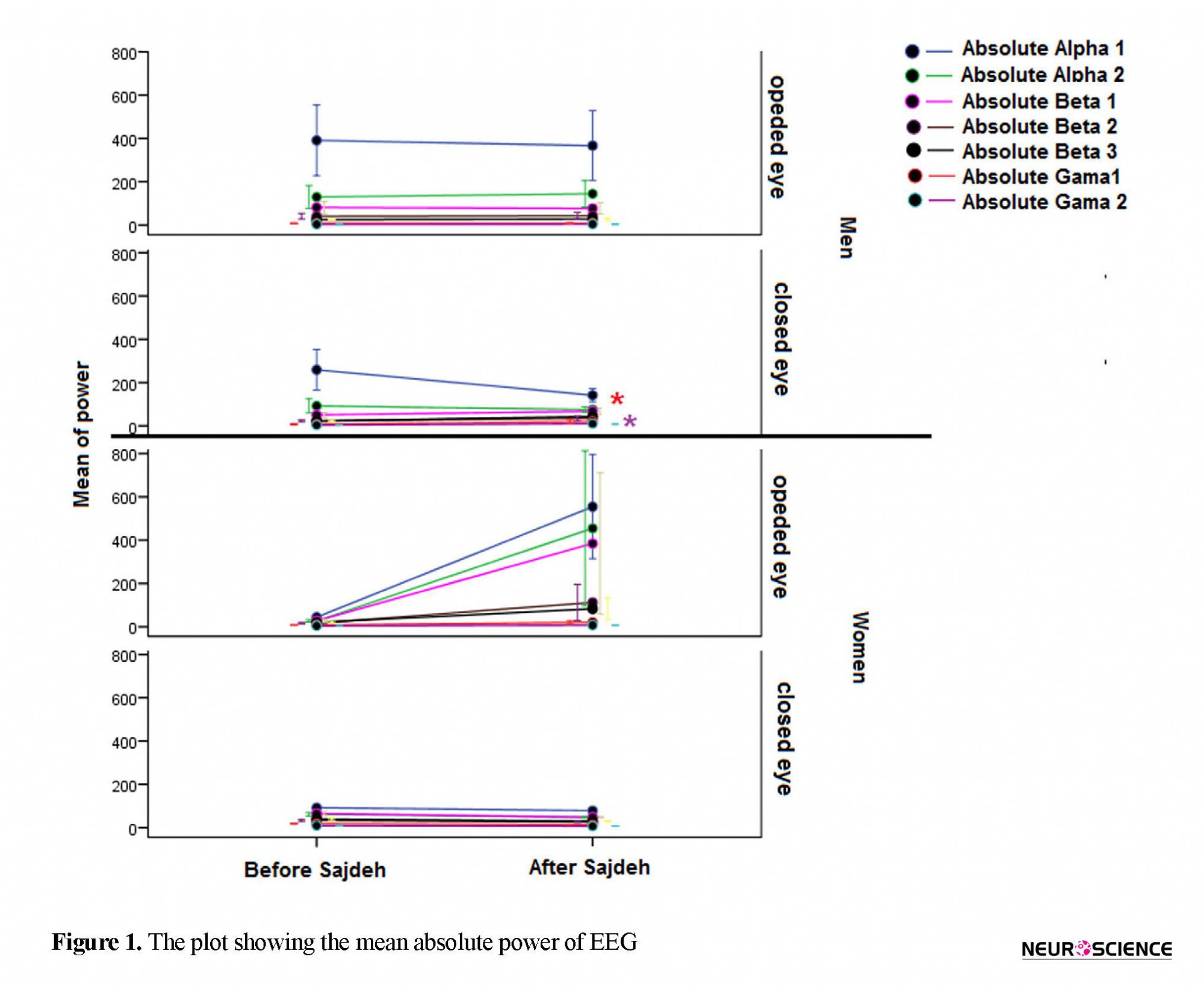
The linear EEG features based on Mann-Whitney U test before and after Sajdah in open and closed eye states are presented in Table 1. The absolute power of band frequency did not show significant difference except an increase of the γ 1 and γ 2 after Sajdah in the state of closed eyes in men (Figure 1). Relative power showed a significant decrease in both states in women. The relative power from β 2 to γ 2 band frequency in the opened eye and from θ to γ 1 band frequency in the closed eye state decreased after Sajdah in women. Unlike women, the relative power band frequency tended to increase after the Sajdah in men in the closed eye state that was significant only in the β 1 band (Figure 2).
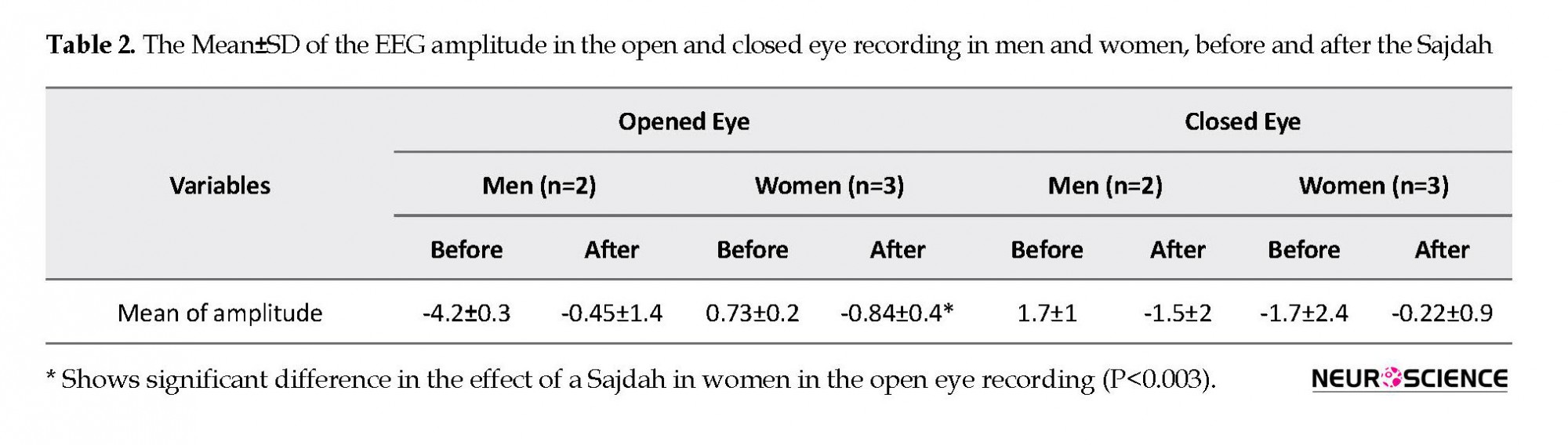
The mean value of EEG amplitude changed after the Sajdah in women in the open eye recordings. But the variation of EEG amplitude did not show significant difference although there was an increasing trend in the women, especially in the open eye recording (Table 2, Figure 3).
• This pilot study assessed the EEG linear and non-linear features of the prefrontal region in the five healthy persons before and 20 seconds after the prostration (Sajdah) position.
• The results showed a significant change in the most relative power band of EEG frequency.
• Non-linear features such as fractal dimension and entropy in time decreased after Sajdah.
• The changes were more significant in women than men.
Plain Language Summary
A 20-second Sajdeh showed a significant change in brain activity of the prefrontal region. It suggests that the complete Muslim praying (Namaz) has several effects that should be studied.
1. Introduction
Prayer is the most important daily duty of Muslims and comprises several acts and positions. During Sajdah, the subject is in prostrating position in the direction of Qibla (the direction that a Muslim prays) and some groups of Muslim community put their forehead on “Mohr”, which is made of dried clay. Rare studies showed the effect of Prayer on bio-signals like Electroencephalography (EEG) and Electrocardiography (ECG).
Doufesh et al. showed a significant decrease in heart rate and sympathetic activity during prayer and especially during Sajdah before and after prayer, especially in actual form (performing Muslim praying with all of nessasary conditions such as Wudu, Gibleh direction, positionings and especial Dhikrs (Doufesh, Ibrahim, Ismail, & Ahmad, 2013; Doufesh, Ibrahim, Ismail, & Wan Ahmad, 2014). They also reported the increase of relative α activity regardless of recitation in comparison with the resting position (Doufesh, Faisal, Lim, & Ibrahim, 2012; Doufesh et al., 2014). It is possible that the increase of α activity and absence of α blocking in the open eye are due to the higher state of calmness and focus as the head touches the ground. The amplitude of the γ band increased after prayer and this effect was significantly higher after listening to music (Ridzwan, Mahmood, Zakaria, & Ali, 2011).
The γ power during actual prayer was statistically higher than just staging prayer positions in the frontal and parietal regions in all stages, especially in the left hemisphere. Increased γ power during prayer, is possibly related to an increase in cognitive and attention processing (Doufesh, Ibrahim, & Safari, 2016).
Usually, prayer is compared with meditation. Many studies showed that some meditations had beneficial effects on the brain (Cahn & Polich, 2006; Ngo, 2013). During meditation, the increase of α band frequency primarily in the frontal region has been demonstrated (Cahn & Polich, 2006). On the other hand, some studies reported adverse effects during and after meditation (Shapiro, 1992). It must be noted that meditation techniques are often done in static posture such as sitting or lying supine, while prayer involves active physical movements.
EEG frequencies are associated with specific functions of the brain as an “electrophysiological signature.” For example, γ oscillations have been related to sensory processing, attention, action selection, conscious awareness and memory, and integrative function (van Wingerden, Vinck, Lankelma, & Pennartz, 2010). Also, δ oscillations have been correlated to motivation, reward processing, memory encoding, retrieval, and learning. The activity of θ band is associated with emotional arousal, fear conditioning and recognition memory (Knyazev, 2007). The α band has been associated with working memory functions and short-term memory. And β oscillations might be associated with the control of cognition (Engel & Fries, 2010; Neuner et al., 2014).
Studies using local field potentials, scalp, or cortical EEG recordings systematically revealed positive correlations between the power of the γ band (>30 Hz) and the BOLD (blood oxygenation level dependent) fluctuations at the same location. They reported the same correlation during cognitive, sensory and motor function at brain regions expected to be activated. The negative correlations exist between the power of the low-frequency ranges (α, β, and θ) and BOLD signals of those regions (Murta, Leite, Carmichael, Figueiredo, & Lemieux, 2015).
The studies about prayer show that the brain activity changes during the Sajdah more than other positions. Then the aim of this pilot study is to determine whether 10 seconds Sajdah as a part of prayer in the Qibla direction could induce any remained changes in the prefrontal area. On the other hand, the biological signals also had complex and chaotic pattern (Kaniusas, 2014) and the non-linear analysis of biological signals was more reliable than linear ones (Melillo, Bracale, & Pecchia, 2011). Then the brain activity was evaluated by non-linear analysis in addition to frequency and amplitude analysis. We hypothesize that Sajdah could change brain activity.
2. Methods
2.1. Study subjects
Two men (aged between 40 and 55 y) and 3 women (aged between 25 and 50 y) participated in the pilot study. We informed them about the study procedure and they signed the approval forms given by the Baqiyatallah University of Medical Sciences. All of the participants had the following characteristics: 1. No history of the psychological disorder based on DSM-5 guideline; 2. No report of surgery or trauma in the cranium and spine regions; 3. No history of taking regular neuropsychological medication; and 4. Always say their prayers regularly on time. They are right handed expect one of the male subjects.
2.2. Data acquisition
EEG signals were obtained from BioMed EEG system 32 channels (Made in Iran). Fp1 and Fp2 electrodes are based on the international 10.20 system attached to the scalp. The reference electrode was put in the Cz position and the ground electrode was attached to the right hand. The skin was cleaned with alcohol before electrode placing to reduce the skin impedance to 20 kΩ or less. Forty seconds of EEG was recorded with eyes open and eyes closed in the resting sitting position before Sajdah and then after 10 seconds of Sajdah in the Qibla direction. The test was done between 6 AM and 8 AM.
2.3. Data processing
EEG data were analyzed offline using MATLAB version R2014b. The obtained data were filtered through 0.2-48 Hz to remove any unwanted artifacts, including EOG (electrooculogram) and EMG (electromyogram). After artifact removal, the data were transformed to the average reference. Twenty-two linear features were extracted from signals in both states (eyes open and closed) before and after the Sajdah. The linear features were the absolute and relative power of frequency bands consisting of θ (4-8Hz), α 1 (8-10 Hz), α 2 (10-12 Hz), β 1 (12-16 Hz), β 2 (16-20 Hz), β 3 (20-30 Hz), γ 1 (30-40 Hz), γ 2 (40-50 Hz) and mean and variance of signal amplitude.
The relative features were calculated using the various band powers divided by the total power of the signal. The non-linear features were approximate entropy, Katz fractal dimension, Petrosian fractal dimension, sample entropy and spectral entropy. The Fp channels were preferred to study because the frontal region is contacted with the ground during the Sajdah.
2.4. Statistical analysis
The non-parametric Mann-Whitney U test was used to compare the EEG features before and after the Sajdah in separate groups in two different states. As a result of the small sample size and similar behavior in Fp1 and Fp2, they were considered together and mean of them was reported.
3. Results
3.1. The linear features of EEG
The linear EEG features based on Mann-Whitney U test before and after Sajdah in open and closed eye states are presented in Table 1. The absolute power of band frequency did not show significant difference except an increase of the γ 1 and γ 2 after Sajdah in the state of closed eyes in men (Figure 1). Relative power showed a significant decrease in both states in women. The relative power from β 2 to γ 2 band frequency in the opened eye and from θ to γ 1 band frequency in the closed eye state decreased after Sajdah in women. Unlike women, the relative power band frequency tended to increase after the Sajdah in men in the closed eye state that was significant only in the β 1 band (Figure 2).
The mean value of EEG amplitude changed after the Sajdah in women in the open eye recordings. But the variation of EEG amplitude did not show significant difference although there was an increasing trend in the women, especially in the open eye recording (Table 2, Figure 3).
3.2. The non-linear features of EEG
The fractal non-linear features of EEG such as Katz fractal dimension and Petrosian fractal dimension showed that the Sajdah decreased significantly the fractal dimension of EEG signals in the prefrontal region in women in open eye state (Figure 4). Approximate entropy and sample entropy decreased significantly in the open eye state in women (Figures 5 and 6).
The Mann-Whitney U test was also used to compare the EEG features between men and women before and after Sajdah. The results showed that gender differences in the EEG features diminished after Sajdah in the open eye state and in the non-linear features in both states of eyes. It means that the trend of changes in the two genders was different after the Sajdah that was significantly evident in the open eye recording (Table 3).
4. Discussion
The aim of this pilot study was to measure the linear and non-linear features of brain activity in the prefrontal region before and after one Sajdah (for 10 seconds), as a part of Muslim daily prayer. In spite of the small sample size, some clearly significant effects of the Sajdah were seen especially in women at the open eye recording. The results showed the decrease of the relative power of bands, especially β and γ oscillations in both open and closed eye recording and decrease of entropy and Petrosian fractal dimension as a non-linear feature of EEG in the open eye state after the Sajdah in women. Increase of the γ power during and after prayer had been demonstrated before. But the participants were men (Doufesh et al., 2016; Ridzwan et al., 2011).
Our study evaluated the women too and showed the opposite effect of one Sajdah to relative γ band in women but not in men. On the other hand, the brain activity after one Sajdah did not cover the effect of prayer and they could be different and not comparable. Therefore the current results confirmed the previous results in men with respect to the absolute power of the γ band.
The non-linear features showed that the entropy and fractal dimension of EEG signals as indices of signal complexity (Ahmadi, Ahmadlou, Rezazade, Azad-Marzabadi, & Sajedi, 2013; Micheloyannis, Vourkas, Bizas, Simos, & Stam, 2003) decreased in women. Ten seconds of Sajdah like any other cognitive tasks (Corsi-Cabrera, Ramos, Guevara, Arce, & Gutierrez, 1993; Kober, Reichert, Neuper, & Wood, 2016) or exposure to electromagnetic field (Papageorgiou, Nanou, Tsiafakis, Capsalis, & Rabavilas, 2004) has inverse effect in brain activity of men and women which could be the result of gender structural differences (Ahmadi et al., 2013; Corsi-Cabrera et al., 1993; Kober et al., 2016; McGivern et al., 1998).
The effects of sex steroids in the human brain may play some role in explaining these differences. The sex steroids interact with neurotransmitters and other hormones such as the oxytocin-vasopressin system in the brain that regulates the brain function (Nguyen, Ducharme, & Karama, 2016). Several studies show that some cognitive abilities are higher in women and some of them are advanced in men (Halpern & LaMay, 2000; Li, 2014; McGivern et al., 1998). The interesting finding shown in Table 3 indicates that the significant baseline linear and non-linear features of EEG between genders decreased or changed after the Sajdah.
The increase of γ power that was seen after the Sajdah has been related to increased activity of the frontal node of the Default Mode Network (DMN), the medial prefrontal cortex (Berkovich-Ohana, Glicksohn, & Goldstein, 2012), and the cognitive activity (van Wingerden et al., 2010). The increase of α oscillation was related to working memory (Neuner et al., 2014) and θ oscillation to emotional processing (Knyazev, 2007). The event-related potentials studies concluded that meditation can increase attention and enhance emotional control that matched with an increase of θ and α oscillations in meditation (Singh & Telles, 2015).
The results of studies in meditation or prayer have been reported in the men or mixed gender group (Cahn & Polich, 2006; Doufesh et al., 2012; Doufesh et al., 2014; Doufesh et al., 2016; Singh & Telles, 2015) and there was no study that measured the effect of meditation or prayer in the brain activity with interaction gender. Whenever the Sajdah as a part of Muslim praying showed some changes but it could not be referred to the effect of the completed prayer.
Some researchers believe that prayer is a type of meditation. But there are significant differences between them in terms of the action and effects. Prayer involves physical movements with specific and fixed saying and pattern but meditation often involves static position without saying in several patterns. The meditation research with expanding the methodological paradigm of cultural setting as the place of meditator, the particular practice, and the state of consciousness of mediators showed several states and trait effects on brain activity, especially increased power of low frequency such as θ and α bands and decrease of γ oscillations over the frontal and midline regions (Berkovich-Ohana et al., 2012; Cahn & Polich, 2006; Kaur & Singh, 2015; Thomas & Cohen, 2014). Whereas prayer increased both the power of α and γ band (Doufesh et al., 2012; Doufesh et al., 2014; Doufesh et al., 2016). Therefore the comparison of them is not always appropriate.
No study has measured non-linear features of EEG in the whole of prayer or meditation or a part of them. Whereas our results showed that Sajdah had a significant effect on the complexity of the EEG signal. The decrease of sample and approximate entropy and Petrosian fractal dimension were detected after the Sajdah in the women, especially in the open eyes state. The signal complexity of EEG is correlated to high-frequency oscillations, especially γ band. (Micheloyannis et al., 2003; Murta et al., 2015; Pravitha, Sreenivasan, & Nampoori, 2005).
On the other hand, the decrease in complexity was shown in inhibition control as an adaptive ability of brain (Huang, Tseng, & Liang, 2015). Lower neural complexity does not always indicate the declines in information processing and cognitive function (McDonough & Nashiro, 2014). The decrease of γ power and signal complexity in women might be the result of shifting of the cognitive processing from sensory and motor processing to inhibition control dominance and decreased activity of the frontal node of DMN. The limitations of the study were the small sample size, limited recorded electrodes, and not recording during Sajdah or in the real prayer and assessment of the whole prayer.
The pilot study showed that 10 seconds of Sajdah in the direction of Qibla while putting the forehead on Mohr significantly influenced the brain activity on the prefrontal region. Sajdah had opposite effects on different genders. In women, the power bands, especially γ and β oscillation and the complexity of signals decreased, especially in open eye recording. Whereas the effect of Sajdah showed an increase in the absolute power of β or γ frequency band of EEG in men.
The genders showed significant baseline differences in the linear and non-linear features of brain activity in the prefrontal region after Sajdah. The findings of the pilot study need to be further evaluated by other studies with enough sample size and in real prayer. The Sajdah is only one part of the prayer which showed significant improvement in brain activity. The Muslims are supposed to do it five times a day. Thus the effect of prayer to maintain and improve brain activity and mental health is critical and needs to be studied more in the future.
Ethical Considerations
Compliance with ethical guidelines
There was no ethical consideration to be considered in this research.
Funding
This research did not receive any specific grant from funding agencies in the public, commercial, or not-for-profit sectors.
Authors' contributions
Boshra Hatef contributed in all parts of the study, conceptualization, methodology, investigation, writing original draft, review and editing. Fateme Yousefzadeh contributed in signal processing and writing the manuscript; Gila Pirzad Jahromi and Ehsan Mokari Manshadi contributed in data gathering, preparing and editing the manuscript.
Conflict of interest
The authors declared no conflict of interest.
Acknowledgments
The authors thank Dr. Reza Khosrowabadi for assistant in signal processing and feature extraction.
References
Ahmadi, K., Ahmadlou, M., Rezazade, M., Azad-Marzabadi, E., & Sajedi, F. (2013). Brain activity of women is more fractal than men. Neuroscience Letters, 535, 7-11. [DOI:10.1016/j.neulet.2012.12.043]
Berkovich-Ohana, A., Glicksohn, J., & Goldstein, A. (2012). Mindfulness-induced changes in gamma band activity-implications for the default mode network, self-reference and attention. Clinical Neurophysiology, 123(4), 700-10. [DOI:10.1016/j.clinph.2011.07.048]
Cahn, B. R., & Polich, J. (2006). Meditation states and traits: EEG, ERP, and neuroimaging studies. Psychological Bulletin, 132(2), 180-211. [DOI:10.1037/0033-2909.132.2.180]
Corsi-Cabrera, M., Ramos, J., Guevara, M. A., Arce, C., & Gutierrez, S. (1993). Gender differences in the EEG during cognitive activity. International Journal of Neuroscience, 72(3-4), 257-64. [DOI:10.3109/00207459309024114]
Doufesh, H., Faisal, T., Lim, K. S., & Ibrahim, F. (2012). EEG spectral analysis on muslim prayers. Association for Applied Psychophysiology and Biofeedback, 37(1), 11-18. [DOI:10.1007/s10484-011-9170-1]
Doufesh, H., Ibrahim, F., & Safari, M. (2016). Effects of Muslims praying (Salat) on EEG gamma activity. Complementary Therapies in Clinical Practice, 24, 6-10. [DOI:10.1016/j.ctcp.2016.04.004] [PMID]
Doufesh, H., Ibrahim, F., Ismail, N. A., & Ahmad, W. A. W. (2013). Assessment of heart rates and blood pressure in different salat positions. Journal of Physical Therapy Science, 25(2), 211-4. [DOI:10.1589/jpts.25.211]
Doufesh, H., Ibrahim, F., Ismail, N. A., & Wan Ahmad, W. A. (2014). Effect of Muslim prayer (salat) on alpha electroencephalography and its relationship with autonomic nervous system activity.Journal of Alternative and Complementary Medicine, 20(7), 558-62. [DOI:10.1089/acm.2013.0426]
Engel, A. K., & Fries, P. (2010). Beta-band oscillations--signalling the status quo? Current Opinion in Neurobiology, 20(2), 156-65. [DOI:10.1016/j.conb.2010.02.015]
Halpern, D. F., & LaMay, M. L. (2000). The smarter sex: A critical review of sex differences in intelligence. Educational Psychology Review, 12(2), 229-46. [DOI:10.1023/A:1009027516424]
Huang, S.-L., Tseng, P., & Liang, W.-K. (2015). Dynamical Change of Signal Complexity in the Brain During Inhibitory Control Processes. Entropy, 17(10), 6834-53. [DOI:10.3390/e17106834]
Kaniusas, E. (2014). Nonlinear behaviour of vital physiological systems. In v. In, P. Longhini (Eds.), International Conference on Theory and Application in Nonlinear Dynamics (ICAND 2012) (pp. 113-21). Berlin: Springer. [DOI:10.1007/978-3-319-02925-2_10]
Kaur, C., & Singh, P. (2015). EEG derived neuronal dynamics during meditation: Progress and challenges. Advances in Preventive Medicine, 2015 (614723), 1-10. [DOI:10.1155/2015/614723]
Knyazev, G. G. (2007). Motivation, emotion, and their inhibitory control mirrored in brain oscillations. Neuroscience & Biobehavioral Reviews, 31(3), 377-95. [DOI:10.1016/j.neubiorev.2006.10.004]
Kober, S. E., Reichert, J. L., Neuper, C., & Wood, G. (2016). Interactive effects of age and gender on EEG power and coherence during a short-term memory task in middle-aged adults. Neurobiology of Aging, 40, 127-37. [DOI:10.1016/j.neurobiolaging.2016.01.015]
Li, R. (2014). Why women see differently from the way men see? A review of sex differences in cognition and sports. Journal of Sport and Health Science, 3(3), 155-62. [DOI:10.1016/j.jshs.2014.03.012]
McDonough, I. M., & Nashiro, K. (2014). Network complexity as a measure of information processing across resting-state networks: evidence from the Human Connectome Project. Frontiers in Human Neuroscience, 8, 409. [DOI:10.3389/fnhum.2014.00409]
McGivern, R. F., Mutter, K. L., Anderson, J., Wideman, G., Bodnar, M., & Huston, P. J. (1998). Gender differences in incidental learning and visual recognition memory: Support for a sex difference in unconscious environmental awareness. Personality and Individual Differences, 25(2), 223-32. [DOI:10.1016/S0191-8869 (98)00017-8]
Melillo, P., Bracale, M., & Pecchia, L. (2011). Nonlinear heart rate variability features for real-life stress detection. Case study: Students under stress due to university examination. BioMedical Engineering OnLine, 10(1), 96. [DOI:10.1186/1475-925X-10-96]
Micheloyannis, S., Vourkas, M., Bizas, M., Simos, P., & Stam, C. J. (2003). Changes in linear and nonlinear EEG measures as a function of task complexity: Evidence for local and distant signal synchronization. Brain Topography, 15(4), 239-47. [DOI:10.1023/A:1023962125598] [PMID]
Murta, T., Leite, M., Carmichael, D. W., Figueiredo, P., & Lemieux, L. (2015). Electrophysiological correlates of the BOLD signal for EEG-informed fMRI. Human Brain Mapping, 36(1), 391-414. [DOI:10.1002/hbm.22623]
Neuner, I., Arrubla, J., Werner, C. J., Hitz, K., Boers, F., Kawohl, W., et al. (2014). The default mode network and EEG regional spectral power: A simultaneous fMRI-EEG study. PLOS One, 9(2), e88214. [DOI:10.1371/journal.pone.0088214]
Ngo, T. L. (2013). [Review of the effects of mindfulness meditation on mental and physical health and its mechanisms of action (French)]. Santé mentale au Québe, 38(2), 19-34. [DOI:10.7202/1023988ar]
Nguyen, T. V., Ducharme, S., & Karama, S. (2017). Effects of sex steroids in the human brain. Molecular Neurobiology, 54(9), 7507-19. [DOI:10.1007/s12035-016-0198-3]
Papageorgiou, C. C., Nanou, E. D., Tsiafakis, V. G., Capsalis, C. N., & Rabavilas, A. D. (2004). Gender related differences on the EEG during a simulated mobile phone signal. NeuroReport, 15(16), 2557-60. [DOI:10.1097/00001756-200411150-00026] [PMID]
Pravitha, R., Sreenivasan, R., & Nampoori, V. P. (2005). Complexity analysis of dense array EEG signal reveals sex difference. International Journal of Neuroscience, 115(4), 445-60. [DOI:10.1080/00207450590520911x] [PMID]
Ridzwan, W. M. F. W. M., Mahmood, N. H., Zakaria, N. A., & Ali, E. A. (2011). Salat and brainwave signal analysis. Journal Teknologi, 54(1), 181-92. [DOI:10.11113/jt.v54.809]
Shapiro, D. H., Jr. (1992). Adverse effects of meditation: A preliminary investigation of long-term meditators. Journal of Psychosomatic Research, 39(1-4), 62-7. [PMID]
Singh, N., & Telles, S. (2015). Neurophysiological effects of meditation based on evoked and event related potential recordings. BioMed Research International, 2015(406261), 1-11. [DOI:10.1155/2015/406261]
Thomas, J. W., & Cohen, M. (2014). A methodological review of meditation research. Frontiers in psychiatry, 5, 74. [DOI:10.3389/fpsyt.2014.00074] [PMID] [PMCID]
van Wingerden, M., Vinck, M., Lankelma, J. V., & Pennartz, C. M. (2010). Learning-associated gamma-band phase-locking of action-outcome selective neurons in orbitofrontal cortex. Journal of Neuroscience, 30(30), 10025-38. [DOI:10.1523/JNEUROSCI.0222-10.2010]
Type of Study: Original |
Subject:
Computational Neuroscience
Received: 2017/08/31 | Accepted: 2018/05/28 | Published: 2019/07/21
Received: 2017/08/31 | Accepted: 2018/05/28 | Published: 2019/07/21
Send email to the article author
| Rights and permissions | |
 |
This work is licensed under a Creative Commons Attribution-NonCommercial 4.0 International License. |
![]()
Copyright © The Author(s);
This is an open access article distributed under the terms of the Creative Commons Attribution License (CC-By-NC), which permits use, distribution, and reproduction in any medium, provided the original work is properly cited and is not used for commercial purposes.
Contact Information
